fetching data ... 
Background: Sjögren’s disease (SjD) is characterized by impaired exocrine glands functions, including reduced salivary flow. Many SjD patients present with lymphocytic infiltrations into the salivary gland often in combination with a shift from physiological IgA plasma cells to autoreactive IgG plasma cells [1,2]. However, lymphocytic infiltrations are frequently absent (focus score = 0) in SjD patients despite minimal salivary flow [3]. Animal experiments indicate that serum IgG from SjD patients can interact with acinar cells and reduce salivary flow by a mechanism dependent on the IgG antibody Fc-part, independently of B- and T-cells [4-6]. Follow-up studies further imply an epitope-dependent reduction in saliva production, possibly related to specific peptides of the SSA/Ro protein (Ro-274)[5,6].
Objectives: While symptom onset, including dryness, frequently precedes the diagnosis of SjD and serum autoantibodies can be detected years before diagnosis [7], the pathogenic properties of antibodies in the context of SjD and salivary gland function are far from being understood, especially in humans. Utilizing a salivary gland organoid (SGO) model, we investigate the pathogenic potential of SjD patient IgG towards salivary glands.
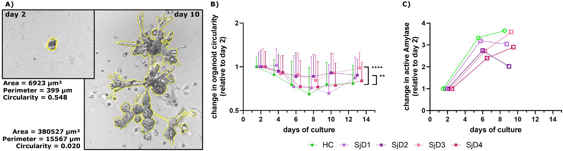
Methods: Salivary gland biopsy material of healthy donors was used to grow SGOs as previously described [8]. Induction of a maturation process leads to growth and extensive branching of the SGO structures (Figure 1A) and upregulation of typical salivary gland genes [9]. Total serum IgG was isolated from 40 SjD patients with salivary gland impairment (unstimulated saliva flow ≤ 0,1 ml/min and stimulated saliva flow ≤ 1 ml/min) and signs of B-cell hyperactivity (serum IgG concentration ≥ 20 g/L and anti-SSA positivity). Control IgG was isolated from the serum of 15 healthy individuals. IgG isolates from 10 SjD patients each and from all 15 healthy individuals were combined into IgG pools. Starting at day 2 of the maturation process, SGOs were exposed to the 4 IgG pools from SjD patients in comparison to the healthy IgG pool, together with interferon-α and active complement proteins. Changes in SGO morphology were monitored over the time of maturation and analyzed with a machine-learning pixel classifier, trained to automatically detect SGO structures. Area, perimeter and circularity were selected as readout parameters, with circularity as a measurement of branching with a value of 1 for perfect circles but approaching 0 for highly-complex structures. Culture supernatant was used in an enzymatic assay to determine the amount of secreted, active α-amylase able to digest polysaccharides.
Results: The 4 different SjD IgG antibodies pools were tested in comparison to the healthy IgG pool for potential interference with SGO maturation. Increase in the perimeter mean of maturing SGO over time was significantly lower in the presence of SjD IgG pool 3 while the total perimeter sum was comparable during the whole maturation timeline. The decrease in mean SGO circularity, i.e. the process of forming branches, was significantly impaired for SjD IgG pools 2 and 3 over the time of observation, especially around day 8 of the SGO culture (Figure 1B). While the relative amount of enzymatically-active α-amylase in the culture supernatant increased for all tested conditions, secretion was impaired in SGOs exposed to SjD IgG pool 2 in comparison to healthy IgG at day 9 of the SGO culture (Figure 1C). Preliminary flow cytometry staining might indicate binding of IgG from SjD patients to the surface of maturing SGOs.
Conclusions: Using an in-vitro organoid system of healthy human salivary glands, we see clear signs of a direct impact of IgG antibodies from some SjD patients to impair the maturation of organoid morphology and function. Since SGO abnormalities were only observed in 2 of 4 SjD IgG pools, presence of pathogenic autoantibodies are likely patient specific in an epitope-dependent manner and could foster identification of a patient subgroup characterized by an antibody-driven pathogenicity of SjD. Our organoid assay might therefore be of special interest in the context of antibody-directed treatments like FcRn-blockers.
REFERENCES: [1] Salomonsson, S. & Wahren‐Herlenius, M. Local production of Ro/SSA and La/SSB autoantibodies in the target organ coincides with high levels of circulating antibodies in sera of patients with Sjögren’s syndrome. Scand. J. Rheumatol. (2003).
[2] Van Ginkel, M. S. et al. Increased Diagnostic Accuracy of the Labial Gland Biopsy in Primary Sjögren Syndrome When Multiple Histopathological Features Are Included. Arthritis Rheumatol. (2024).
[3] Mossel, E. et al. Histopathology, salivary flow and ultrasonography of the parotid gland: three complementary measurements in primary Sjögren’s syndrome. Rheumatology (2022).
[4] Robinson, C. P. et al. Transfer of human serum IgG to nonobese diabetic Igμnull mice reveals a role for autoantibodies in the loss of secretory function of exocrine tissues in Sjögren’s syndrome. Proc. Natl. Acad. Sci. (1998).
[5] Scofield, R. H., Asfa, S., Obeso, D., Jonsson, R. & Kurien, B. T. Immunization with Short Peptides from the 60-kDa Ro Antigen Recapitulates the Serological and Pathological Findings as well as the Salivary Gland Dysfunction of Sjögren’s Syndrome. J. Immunol. (2005).
[6] Maier-Moore, J. S. et al. Passive transfer of antibodies to the linear epitope 60 kD Ro 273–289 induces features of Sjögren’s syndrome in naive mice. Clin. Exp. Immunol. (2015).
[7] Theander, E. et al. Prediction of Sjögren’s Syndrome Years Before Diagnosis and Identification of Patients With Early Onset and Severe Disease Course by Autoantibody Profiling. Arthritis Rheumatol. (2015).
[8] Pringle, S. et al. Salivary Gland Stem Cells Age Prematurely in Primary Sjögren’s Syndrome. Arthritis Rheumatol. (2019).
[9] Yang, T. et al. Cell Composition Analysis of Matched Salivary Organoid and Adherent Cultures: Choose Your Sjögren Disease Research Tool Carefully. J. Rheumatol. (2024).
Acknowledgments: NIL.
Disclosure of Interests: None declared.