fetching data ... 
Background: The synovial fluid (SF) of rheumatoid arthritis (RA) patients contains a unique subset of SPP1+ macrophages, which play a significant role in the pathogenesis of RA by inducing the activation of fibroblast-like synoviocytes (FLS). Based on single-cell RNA sequencing (scRNA-seq) data from the SF, ST, and peripheral blood of the same RA patient, SPP1+ macrophages are specifically enriched in the SF and ST of the joint, but are virtually absent in peripheral blood, which limits the in-depth study of these cells. Previous research of SPP1+ macrophages has predominantly focused on RA patients, with a gap in understanding the distribution and function of Spp1+ macrophages in animal models.
Objectives: This study aims to explore the distribution and functional characteristics of Spp1+ macrophages in the joints of mice by constructing a collagen-induced arthritis (CIA) mouse model. This approach addresses the limitations of RA clinical sample research, and provides crucial experimental evidence for the study of RA pathogenesis and the development of therapeutic strategies.
Methods: The CIA mouse model was established, and scRNA-seq was performed on the joints of CIA and control mice. After quality control, the sequencing data was annotated and clustered to identify cell populations. Flow cytometry was used to analyze the proportion of Spp1+ macrophages in the peripheral blood, spleen, and joints of CIA mice based on previously established gating strategies. Micro-computed tomography (Micro-CT) was used to assess bone metabolism-related parameters in mice, and their correlation with the proportion of Spp1+ macrophages was analyzed.
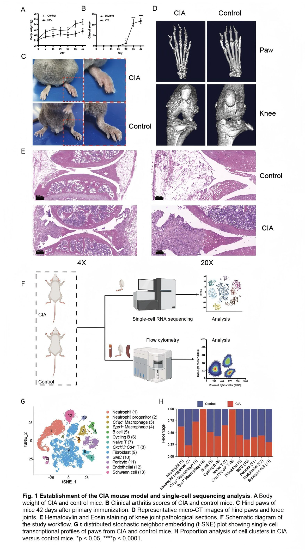
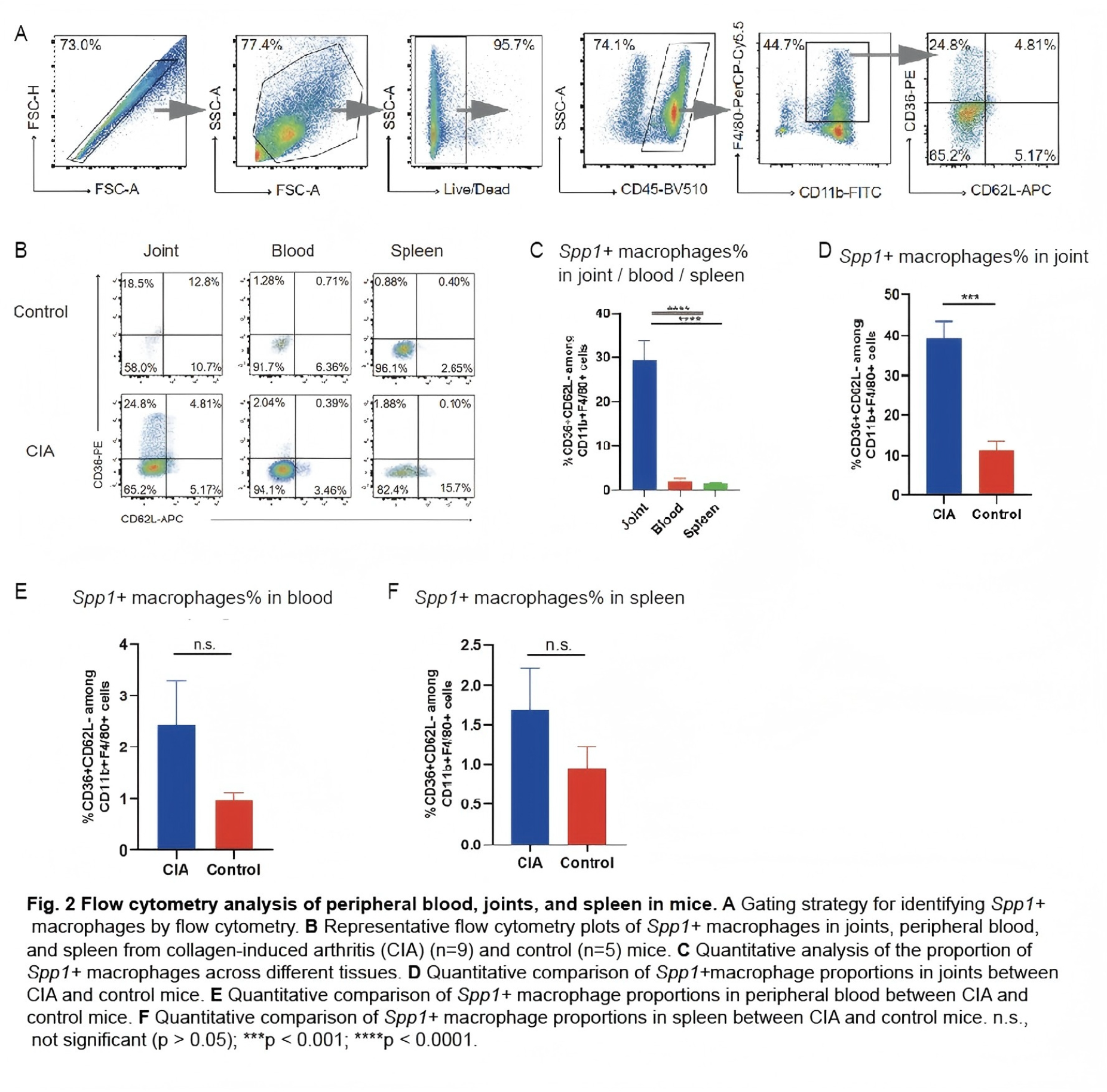
Results: Following the classic protocol for CIA mouse model induction, on day 42 post-primary immunization, CIA mice exhibited significant weight loss compared to controls, along with more severe clinical scores, synovial inflammation, and joint destruction, confirming successful model establishment (Figure 1A-E). Single-cell RNA sequencing of the paws from one CIA mouse and one control mouse revealed the specific presence of Spp1+ macrophages in the swollen joints of CIA mice, which were absent in controls (Figure 1F-H). Further validation of the distribution of pathogenic Spp1+ macrophages was conducted using flow cytometry (Figure 2A). The proportion of Spp1+ macrophages was significantly higher in the joints than in peripheral blood and spleen (Figure 2B-C). Moreover, the proportion of Spp1+ macrophages in the joints of CIA mice was significantly elevated compared to controls, while no such differences were observed in peripheral blood and spleen (Figure 2E-F). Three-dimensional reconstruction of mouse knee joints showed severe joint surface damage and bone erosion in CIA mice. Assessment of four bone-related parameters in the proximal tibia, namely bone volume (BV)/tissue volume (TV), bone surface (BS)/TV, trabecular number (Tb.N), and trabecular sepamtion (Tb.Sp), revealed significant decreases in BV/TV, BS/TV, and Tb.N in the CIA group (not presented), while Tb.Sp, an indicator of osteoporosis severity, was higher, indicating bone loss and osteoporosis in CIA mice. Spearman correlation analysis revealed that the proportion of Spp1+ macrophages in the joints was significantly negatively correlated with BV/TV, BS/TV, and Tb.N but positively correlated with Tb.Sp. These findings suggest that the increased proportion of Spp1+ macrophages in the joints may be associated with reduced bone volume, decreased trabecular number, and increased osteoporosis in CIA mice.
Conclusions: This study reveals that Spp1+ macrophages are specific enrichment in CIA mouse joints and their association with abnormal bone metabolism. These findings offer new insights into RA pathogenesis, address clinical sample limitations, and provide experimental evidence for developing therapeutic strategies.
REFERENCES: [1] Xia, X. et al. Single cell immunoprofile of synovial fluid in rheumatoid arthritis with TNF/JAK inhibitor treatment. Nat Commun 16, 2152, doi:10.1038/s41467-025-57361-0 (2025).
Acknowledgments: NIL.
Disclosure of Interests: None declared.