fetching data ... 
Background: Antinuclear antibodies (ANA) are established biomarkers for the diagnosis and monitoring of autoimmune diseases. Their detection and pattern differentiation commonly rely on indirect immunofluorescence (IIF) on human epithelial type 2 (HEp-2) cells, with fluorescence patterns visually assessed by diagnosticians [1]. However, these approaches have inherent limitations, including observer dependence and restricted resolution, and cannot resolve the fact that different ANA staining patterns may occur within the same autoimmune disease, while identical patterns can be shared across distinct diseases. To overcome these limitations, super-resolution imaging combined with automated analysis is essential for capturing and interpreting the complexity of ANA patterns. High-resolution images provide significantly more detail than standard fluorescence imaging, enabling easier identification and potentially facilitating the discovery of novel ANA patterns.
Objectives: We aimed to assess whether super-resolution stimulated emission depletion (STED) microscopy [2] enhances automated ANA pattern classification compared to conventional fluorescence microscopy. Using two-dimensional images acquired from both methods, we developed machine learning models to classify ANA patterns according to the International Consensus on Antinuclear Antibody Patterns (ICAP) and compared the classification accuracy between fluorescence-based and STED-based models.
Methods: Immunofluorescence ANA staining was carried out according to the manufacturer’s (Euroimmun, Lübeck, Germany) standard protocol, except that a STAR RED 647–conjugated secondary antibody (Abberior, Göttingen) was used instead of fluorescein isothiocyanate (FITC). Confocal images were acquired on an Abberior FACILITY Line microscope (Abberior Instruments GmbH, Germany) operated in standard point-scanning mode. Samples were imaged using a 40×/1.3 NA oil-immersion objective with the 640-nm excitation laser; the 775-nm STED depletion beam was disabled. Laser power and detector gain were optimized to minimize photobleaching while maintaining sufficient signal. Image acquisition and basic corrections were performed using the Lightbox control software, and images were processed in Imspector. Super-resolution images were acquired using an Abberior FACILITY Line STED microscope (Abberior Instruments GmbH, Germany). Samples were labeled with Abberior STAR RED, fixed, and mounted in a refractive index–matched medium according to the manufacturer’s protocol. Imaging was performed using a 40×/1.3 NA oil-immersion objective in STED mode. The excitation laser operated at 640 nm, while the depletion (STED) laser operated at 775 nm, typically set to 40–60% of its maximum output power to balance resolution and minimize photobleaching. Images were acquired in point-scanning mode with time-gated detection to suppress background fluorescence. System calibration and drift correction were performed automatically using the FACILITY Line control software (Lightbox). Image reconstruction and processing were carried out using Imspector software. Almost all of the images formed pairs showing the same sample captured with both microscopy techniques. The staining patterns were established and classified according to the ICAP, which currently recognizes 32 HEp-2 IIFA patterns designated AC-0 to AC-31. Classification was performed by diagnosticians with expertise in recognizing both competent- and expert-level ICAP patterns. The machine learning data pipeline was developed in Python using the TensorFlow and Keras frameworks.
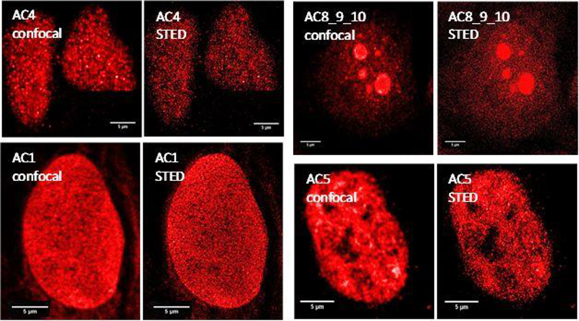
Results: Our ANA image dataset included both confocal and STED images labeled according to ICAP classes: AC1 (homogeneous), AC2 (dense fine speckled), AC3 (centromere), AC4 (fine speckled), AC5 (large speckled), and AC8 (nucleolar homogeneous) (Figure 1). In total, the dataset comprised 2,920 fluorescence and 2,907 STED images, cropped to single nuclei and balanced via data augmentation. Figure 1. shows the comparison of confocal and STED images of identical ANA patterns. STED microscopy provides enhanced resolution, revealing finer structural details that are not visible in conventional confocal images. We developed machine learning models using transfer learning with pretrained EfficientNet and ResNet architectures, applying optimized classification heads and fine-tuning. The best performance was achieved with EfficientNetB0, reaching 94.8% accuracy for confocal images and 87.8% for STED images. Although STED provides higher-resolution imaging of ANA patterns, classification accuracy was lower, likely due to misalignment with current ICAP categories and the complexity of STED detail. This dataset represents the first published collection of ANA images acquired by STED microscopy and establishes a foundation for future research on ANA pattern classification.
Conclusions: We present the first dataset of ANA images acquired using STED super-resolution microscopy. While classification models trained on STED images performed worse than those based on confocal images, this likely reflects limited training data rather than an inherent limitation of STED. The high information density of super-resolution images demands substantially larger datasets to fully leverage their potential in automated pattern recognition. STED microscopy offers a major advance for ANA diagnostics by revealing fine structural details that may correspond to previously unrecognized pattern variants. Future studies with expanded STED datasets are needed to determine whether super-resolution imaging can enable discovery of novel ANA subcategories. Combining STED with nanobody-based labeling is recommended to achieve precise visualization of intracellular targets and facilitate detailed three-dimensional modeling of ANA antigen distribution
Comparison of representative confocal and STED images of ANA patterns in the dataset.
REFERENCES: [1] Bossuyt X et al. Clin Chim Acta. 2013 Jan 16;415:101-6.
[2] Willig KI et al. Nat Methods. 2007 Nov;4(11):915-8.
Acknowledgments: NIL.
Disclosure of Interests: None declared.