fetching data ... 
Background: The emergence of immune-related adverse events (irAEs) poses a major challenge to the advances in immune checkpoint inhibitors (ICIs) therapy, necessitating urgent breakthroughs in predictive diagnostics. However, the dynamics of peripheral immune cells during the development of irAEs remain incompletely characterized, underscoring the need to elucidate their temporal patterns to uncover immune disturbances and identify biomarkers.
Objectives: We performed longitudinal clustering analysis of prospectively collected peripheral blood samples from patients undergoing ICIs therapy, aiming to identify biomarkers for predicting the occurrence, severity and organ involvement of irAEs.
Methods: In this prospective study, patients with lung cancer receiving ICIs were enrolled at Peking Union Medical College Hospital and were consecutively followed up for the development of irAEs. Peripheral blood samples were collected at baseline (T0), early treatment (T1, 1-3 weeks), late treatment (T2, 3-6 months), and at the onset of irAE (Tae). Comprehensive immune profiling was performed using multicolor flow cytometry. We utilized Mfuzz clustering analysis to characterize immune cell trajectories and calculated the change in cell frequencies (ΔT) from T0 to subsequent time points (ΔTae, ΔT1, and ΔT2) to identify time-dependent immune signatures predictive of irAE occurrence, severity, and specific organ involvement.
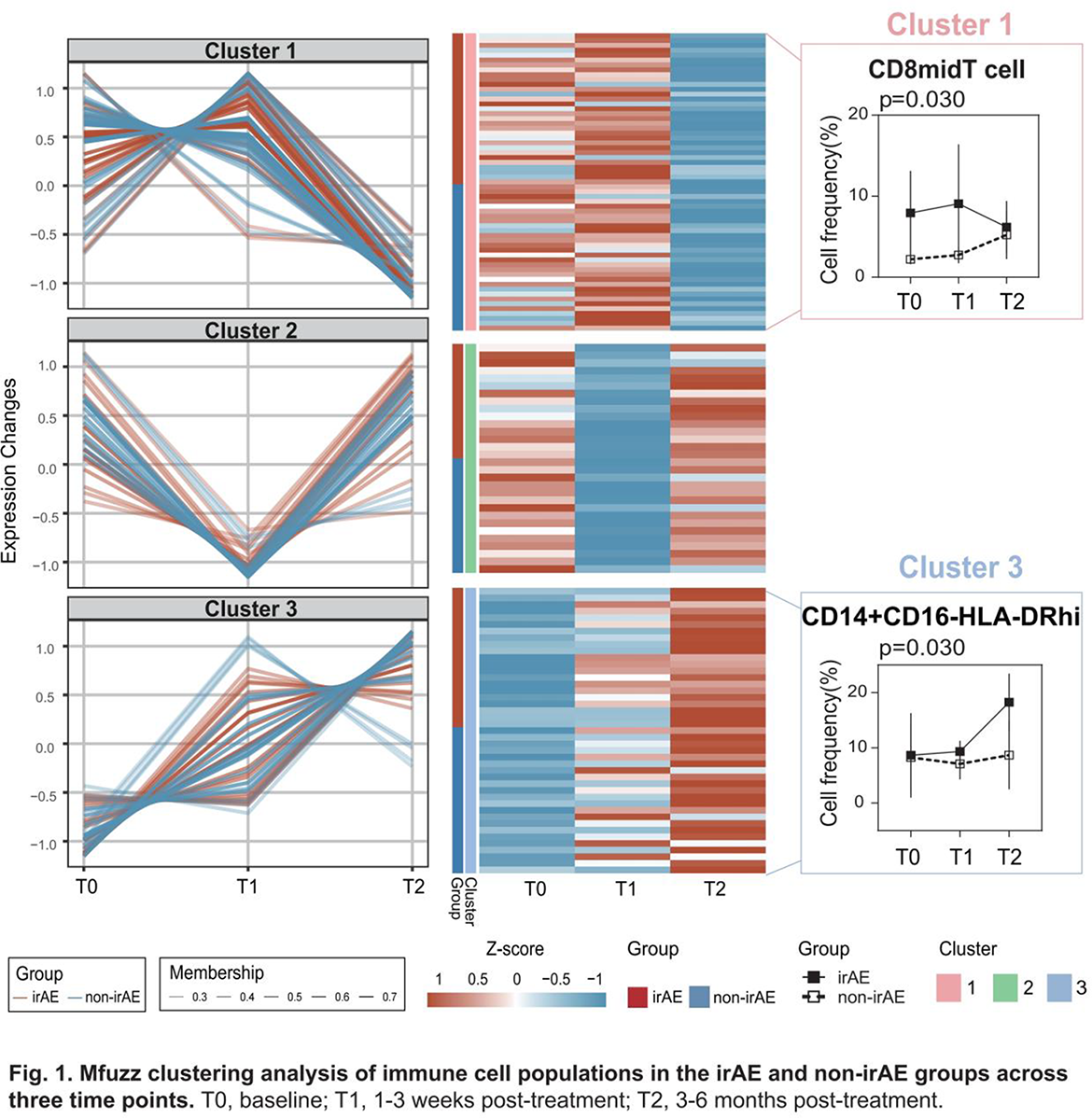
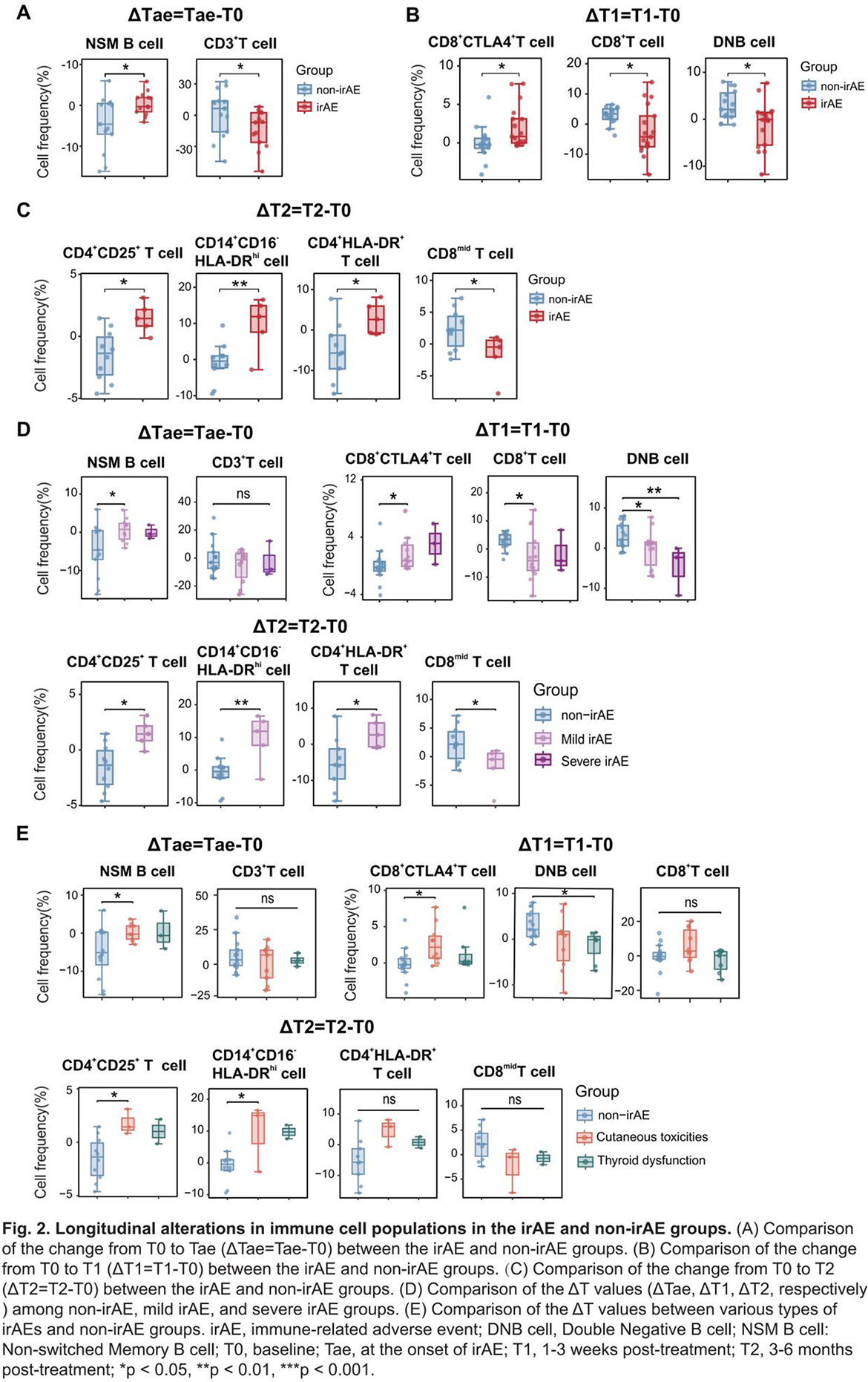
Results: Among the 60 lung cancer patients who received ICIs, 26 (43.3%) developed irAEs. Mfuzz clustering highlighted the distinct dynamics of CD8 mid T cells and CD14 + CD16 - HLA-DR hi monocytes during ICIs therapy ( Figure 1 ). At ΔTae, patients with irAEs exhibited a significant expansion of non-switched memory (NSM) B cells and a reduction in CD3 + T cells, whereas non-irAE patients showed opposite trends ( Figure 2A ). During early treatment (ΔT1), the irAE group showed a greater increase in CD8 + CTLA-4 + T cells and greater reductions in both total CD8 + T cells and double-negative B (DNB) cells ( Figure 2B ). At ΔT2, significantly greater alterations were observed in CD4 + CD25 + T cells, CD4 + HLA-DR + T cells, CD14 + CD16 − HLA-DR hi monocytes, and CD8 mid T cells within the irAE group ( Figure 2C ). Stratified analysis confirmed that the changes (ΔT) in NSM B cells, CD8 + CTLA-4 + T cells, DNB cells, CD4 + CD25 + T cells, and CD14 + CD16 − HLA-DR hi monocytes correlated with both clinical severity and specific organ involvement of irAEs ( Figure 2D-E ).
Conclusions: In conclusion, our study highlights the dynamic immune remodeling during ICIs therapy, identifying unique cellular signatures and their predictive value for the occurrence, severity, and organ involvement of irAEs. These findings underscore the importance of longitudinal monitoring of immune cell subsets to improve risk stratification and management of irAEs.
REFERENCES: NIL.
Acknowledgments: NIL.
Disclosure of Interests: None declared.