fetching data ... 
Background: Ligament ossification disorders represent a significant clinical challenge, encompassing conditions such as ankylosing spondylitis, spinal stenosis, and post-traumatic heterotopic ossification. These diseases involve the pathological transformation of soft connective tissues into ectopic bone, leading to chronic pain, reduced mobility, and neurological compromise. Despite their clinical impact, the fundamental molecular and cellular drivers of this aberrant ossification process remain incompletely understood. Current research has primarily focused on dysregulated osteogenic differentiation pathways and inflammatory mediators. However, the potential contributions of specific metabolic reprogramming events and downstream signaling modifications within the ligament microenvironment are largely unexplored. A deeper understanding of these underlying mechanisms is crucial for developing novel diagnostic and therapeutic strategies to prevent or reverse pathological bone formation.
Objectives: This study aimed to systematically elucidate the key cellular populations and core molecular mechanisms driving ligament heterotopic ossification. By integrating multi-omics analyses of clinical samples with high-throughput functional screening, we sought to identify the critical pathogenic cell subsets and dysregulated biological pathways within the pathological microenvironment. These findings were further validated through in vitro and in vivo functional experiments. Ultimately, this research seeks to provide a novel theoretical foundation for understanding the pathogenesis of ligament ossification and to uncover potential therapeutic targets for its intervention.
Methods: Single-cell RNA sequencing was performed on ossified spinal ligament samples from patients and non-ossified trauma controls to map the cellular landscape and identify dysregulated pathways. Candidate genes from this discovery phase were then subjected to a CRISPR-Cas9-mediated pooled overexpression screen in a murine tendon progenitor cell line, where a multiplex phenotypic assay evaluated their impact on proliferation, osteogenic differentiation, mitochondrial function, and gene expression. The functional role of the prioritized pathway was further investigated in vivo using a mouse model of patellar tendon defect-induced heterotopic ossification, coupled with pharmacological activation or inhibition. The relevance of the downstream protein modification O-GlcNAcylation was assessed across human disease samples and additional animal models via immunofluorescence, and its functional necessity was tested by pharmacologically manipulating its levels both in cultured ligament cells and in the mouse model.
Results: Single-cell transcriptomic profiling of human ossified ligaments identified a distinct pathogenic fibroblast-like subpopulation (CRTAC1 + ) co-enriched for osteogenic programs and aberrantly activated nucleoside sugar metabolism, including genes such as GFPT2 , UAP1 , and UGDH . A subsequent integrative functional screen using CRISPR-mediated overexpression pinpointed core enzymes of the hexosamine biosynthesis pathway (HBP), particularly Gfpt2 and Uap1 , as key regulators promoting ossification in progenitor cells by enhancing extracellular matrix production, mitochondrial energetic capacity, and the expression of osteogenic markers. In a mouse model of trauma-induced patellar tendon heterotopic ossification, pharmacological activation of the HBP pathway significantly increased ectopic bone volume and tissue maturity, whereas its inhibition markedly suppressed the ossification process. This regulatory effect was functionally coupled to the levels of the HBP-dependent protein modification O-GlcNAcylation at the lesion site. Elevated O-GlcNAcylation was further established as a conserved molecular feature across diverse human ligament ossification disorders (ankylosing spondylitis and lumbar spinal stenosis) and in periarticular ectopic bone in a separate inflammatory arthritis model. Functional experiments directly modulating O-GlcNAcylation levels confirmed its pro-ossification role.
Conclusions: This study delineates a novel pathogenic axis linking cellular metabolic reprogramming to pathological bone formation in ligament ossification. We demonstrate that the hexosamine biosynthesis pathway (HBP) is aberrantly activated and functionally drives the ossification process. Critically, this effect is primarily mediated through its downstream protein O-GlcNAcylation. Functional experiments confirm that the levels of this modification causally regulate osteogenic differentiation in ligament cells and ectopic bone formation in vivo. Importantly, elevated O-GlcNAcylation represents a conserved molecular hallmark across ligament ossification disorders of distinct etiologies. These findings establish the HBP/O-GlcNAcylation axis as a central regulatory mechanism and a promising therapeutic target for preventing or treating pathological ligament ossification.
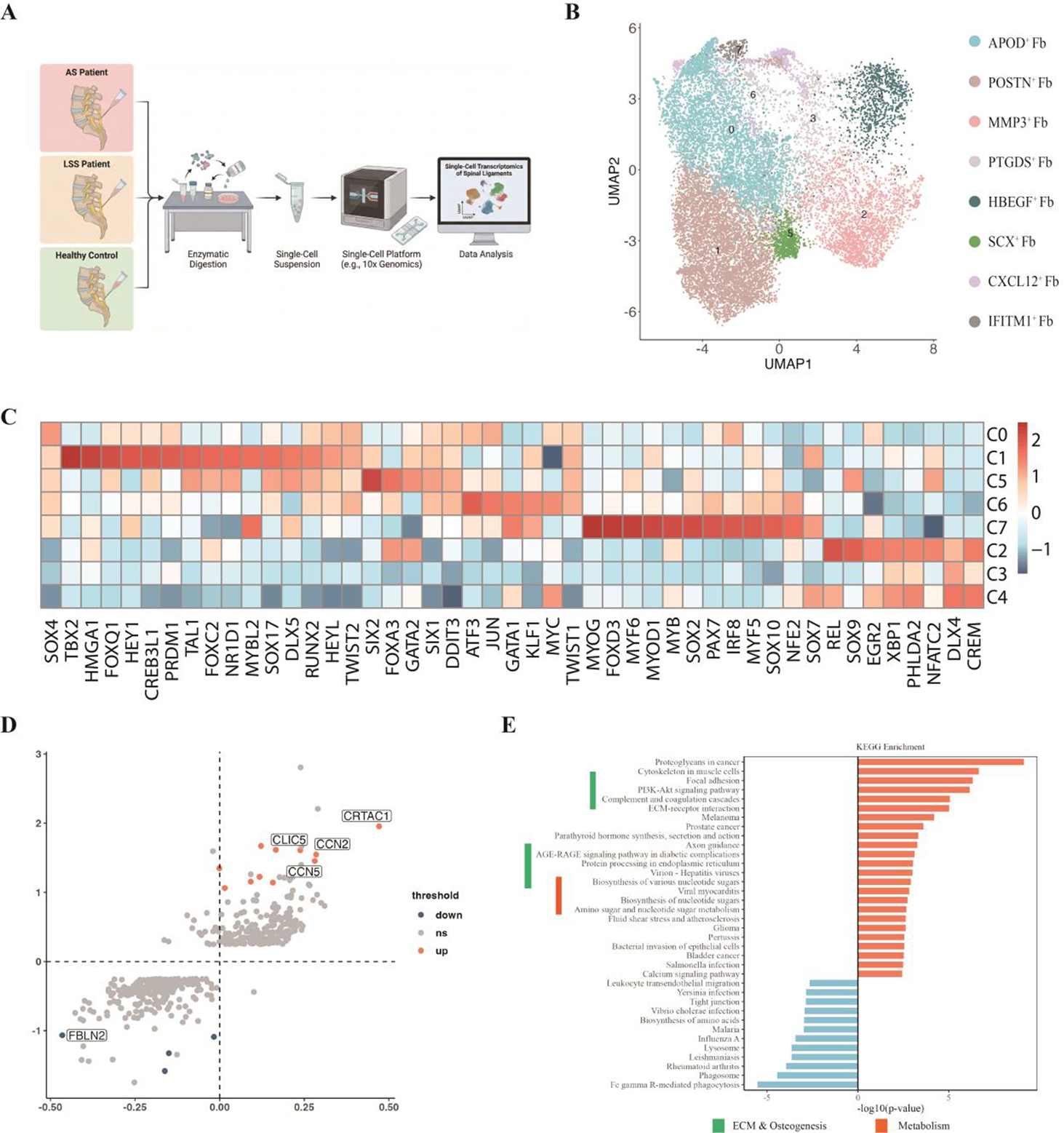
A) Workflow of spinal ligament sample collection, processing, and single-cell analysis. Figure1B) Single-cell visualization of spinal ligament fibroblasts by UMAP. Figure1C) Heatmap of marker gene expression across fibroblast subpopulations. Figure1D) Volcano plot of differentially expressed genes in ligament fibroblasts from ectopic ossification disease versus healthy controls. Figure1E) Pathway enrichment analysis of differentially expressed genes in CRTAC1 + ligament fibroblasts.
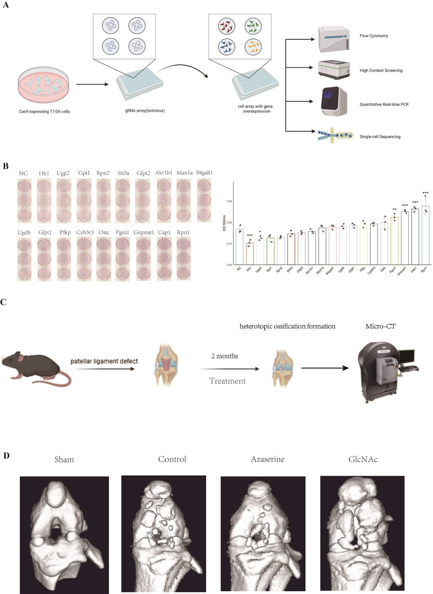
A) Schematic workflow of CRISPR-Cas9 screening coupled with phenotypic screening in tendon stem/progenitor TT-D6 cells. Figure2B) SAlizarin Red S staining and quantification of TT-D6 cells overexpressing different genes after osteogenic induction. Figure2C) Schematic workflow of the mouse patellar tendon defect-induced ectopic ossification model and micro-CT analysis. Figure2D) Representative micro-CT images of patellar tendon defect model with various treatments.
REFERENCES: NIL.
Acknowledgments: NIL.
Disclosure of Interests: None declared.