fetching data ... 
Background: Recent thymic emigrants (RTEs) represent the youngest naïve T-cell output and provide a sensitive readout of early T-cell programming, yet their contribution to rheumatoid arthritis (RA) remains poorly resolved because RTE phenotypes and functions can be reshaped in inflammatory settings [1,2]. Emerging evidence suggests that even very early T-cell populations in RA may exhibit activation and metabolic deviations [3].
Objectives: To quantify CD4 and CD8 RTE alterations in RA across inflammatory arthritides, define their functional and metabolic remodeling, map their integration into immune interaction networks, and assess their translational value as biomarkers of treatment response and remission.
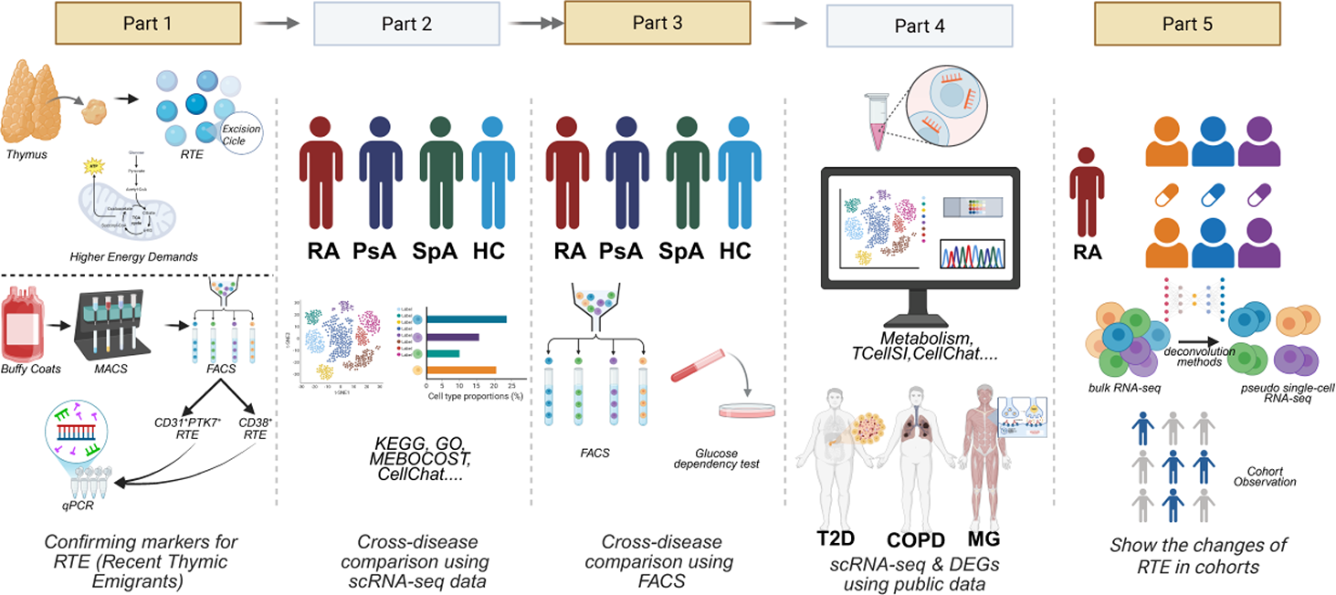
Methods: RTEs were quantified and phenotyped by multicolor flow cytometry, with thymic recency supported by T-cell receptor excision circle (TREC) measurements [2]. Single-cell profiling across Healthy controls, RA, psoriatic arthritis (PsA) and spondyloarthritis (SpA) was used to resolve RTE state programs and metabolic features. Functional metabolic dependencies were confirmed using a translation-based single-cell metabolic assay [4]. Intercellular communication and metabolite-linked signaling programs were reconstructed to position RTEs within systemic immune networks [5,6]. Translational relevance was tested in independent RA therapy cohorts and longitudinal datasets to evaluate whether RTE-derived signatures track clinical improvement and remission.
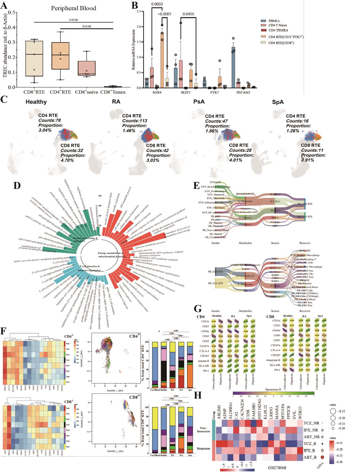
Results: RTEs exhibited high TREC content and SOX4 expression(Figure2A&2B), supporting recent thymic origin. RA showed the highest absolute numbers of circulating CD4 and CD8 RTEs(Figure2C), yet a reduced proportional representation within the naïve pool compared with Healthy controls and PsA, indicating a reshaped naïve compartment rather than simple depletion. RA RTEs displayed coordinated functional remodeling(Figure2D,F), including increased quiescence and senescence-associated programs together with cytotoxic and progenitor-exhaustion–like features. In parallel, RA RTEs demonstrated broad immunometabolic activation involving glycolysis, fatty-acid metabolism, OXPHOS/TCA and PPP pathways, supported by functional metabolic dependency profiling [4]. Network analyses identified RTEs as prominent interaction hubs engaging multiple immune compartments, with key axes including MIF–CD74/CD44, APP–CD74 and CD86–CD28 signaling [5]. Metabolite-linked communication highlighted RA-associated crosstalk involving glutamine, prostaglandin E2 and D-mannose(Figure2E,G) [6]. Importantly, RTE states were therapeutically modifiable: TNF blockade (infliximab) preferentially expanded a metabolically active CD4 RTE compartment, whereas methotrexate more strongly increased CD8 RTE representation. A 15-gene protein-coding RTE signature tracked longitudinal DAS28 improvement and consistently distinguished responders from non-responders and remission from non-remission across multiple cohorts, supporting its translational utility(Figure2H).
Conclusions: RA is associated with quantitative expansion and functional reprogramming of RTEs, positioning thymus-recent cells as active nodes within metabolic–inflammatory immune networks. The therapy-specific modulation of CD4 versus CD8 RTE compartments and the robustness of an RTE-derived signature across independent cohorts support RTEs as clinically actionable biomarkers for monitoring response and remission, and highlight early naïve T-cell programming as a tractable layer for immune modulation in RA.
REFERENCES: [1] Haines CJ, Giffon TD, Lu LS, et al. Human CD4+ T cell recent thymic emigrants are identified by protein tyrosine kinase 7 and have reduced immune function. J Exp Med 2009;206:275–285. doi:10.1084/jem.20080996.
[2] Hazenberg MD, Otto SA, Cohen Stuart JW, et al. T cell receptor excision circles as markers for recent thymic emigrants: basic aspects, technical approach, and guidelines for interpretation. J Mol Med 2001;79:631–640.
[3] Kraus FV, Eckstein V, Klotz LV, et al. AB0025 Peripheral blood CD4+ and CD8+ recent thymic emigrants in rheumatoid arthritis and psoriatic arthritis patients display an activated phenotype. Ann Rheum Dis 2022;81(Suppl 1):1147. doi:10.1136/annrheumdis-2022-eular.2351.
[4] Argüello RJ, Combes AJ, Char R, et al. SCENITH: A flow cytometry-based method to functionally profile energy metabolism with single-cell resolution. Cell Metab 2020;32:1063–1075.e7. doi:10.1016/j.cmet.2020.11.007.
[5] Jin S, Guerrero-Juarez CF, Zhang L, et al. Inference and analysis of cell-cell communication using CellChat. Nat Commun 2021;12:1088. doi:10.1038/s41467-021-21246-9.
[6] Zheng R, et al. MEBOCOST maps metabolite-mediated intercellular communications using single-cell RNA-seq. Nucleic Acids Res 2025;53:gkaf569.
Acknowledgments: NIL.
Disclosure of Interests: None declared.