fetching data ... 
Background: Behçet’s syndrome (BS) is a systemic autoimmune vasculitis capable of affecting vessels of all sizes, often leading to multi-organ involvement [1]. Large-artery involvement represents a particularly severe phenotype, associated with poor prognosis and elevated mortality [2]. High-resolution multi-omics approaches offer a powerful, unbiased platform for characterizing disease mechanisms at cellular and molecular levels [3], enabling a comprehensive exploration of the pathogenesis of BS with major artery involvement.
Objectives: In this study, we aim to explore the pathogenesis of BS with large artery involvement through integrated single-cell transcriptomics and proteomics.
Methods: We enrolled six patients with recurrent aortic-involved BS and four healthy controls (HCs) for single-cell transcriptomic sequencing of peripheral blood mononuclear cells (PBMCs). In addition, we analyzed CD8 + T-cell counts in peripheral blood from 201 BS patients and 114 HCs using flow cytometry. Furthermore, serum proteomic profiling was performed in 16 patients with large-artery involvement and 16 matched healthy controls.
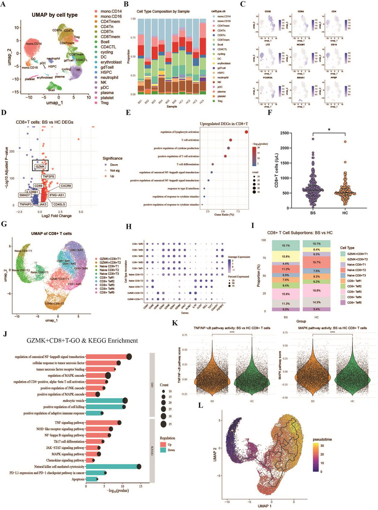
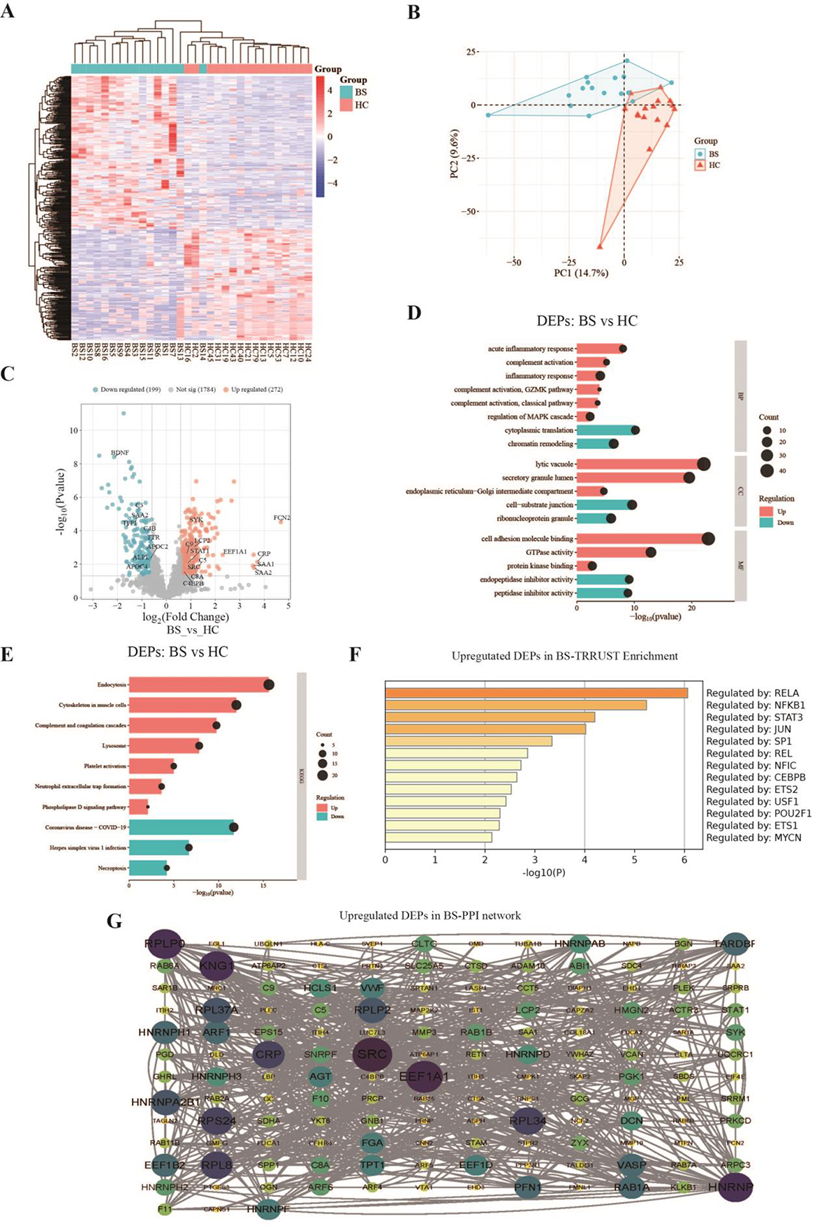
Results: We classified PBMCs from patients with BS and HCs into distinct cell subsets through single-cell transcriptomics and performed differential gene expression analysis. (Figure1 A, B, C) The results revealed a significant upregulation of the GZMK gene in CD8 + T cells from BS patients, with differentially expressed genes enriched in multiple inflammatory signaling pathways. (Figure1 D, E) Flow cytometry further confirmed that the proportion of peripheral blood CD8 + T cells was significantly higher in BS patients than in HCs. (Figure1 F) Subsequent clustering analysis of CD8 + T cells demonstrated an elevated proportion of GZMK + CD8 + T cells in the BS group. (Figure1 G, H, I) Compared to other CD8 + T-cell subsets, GZMK + CD8 + T cells exhibited enhanced activity in inflammatory pathways such as NF-κB and MAPK, alongside reduced cytotoxic function. (Figure1 J) Gene-set scoring also indicated higher NF-κB and MAPK pathway activity in CD8 + T cells from BS patients. (Figure1 K) Pseudotime trajectory analysis suggested that GZMK + CD8 + T cells represent an intermediate differentiation stage within the CD8 + T-cell lineage. (Figure1 L) Serum proteomic profiling identified 272 upregulated and 199 downregulated proteins in BS patients compared to HCs. (Figure2 A, B, C) These differentially expressed proteins were enriched in MAPK cascade and complement activation pathways, including those associated with GZMK. (Figure2 D, E) TRRUST enrichment analysis indicated that the upstream regulators of the upregulated proteins were linked to the NF-κB pathway. (Figure2 F) Protein-protein interaction network analysis highlighted SRC as the most highly connected hub among the upregulated proteins. (Figure2 G).
Conclusions: Patients with arterial-involved BS exhibit elevated levels of GZMK + CD8 + T cells in peripheral blood. GZMK + CD8 + T cells may contribute to autoimmune damage in BS patients through activation of the NF-κB and MAPK pathways, as well as complement signaling.
(A ) UMAP visualization of cell clusters in PBMCs from 6 vascular-type BS patients and 4 HCs. (B ) Proportion of each cell cluster relative to total PBMCs in the 6 BS patients and 4 HCs. (C ) UMAP visualization showing expression of classical cell markers in PBMCs from 6 BS patients and 4 HCs. (D ) Volcano plot of differentially expressed genes in peripheral blood CD8 + T cells between BS patients and HCs; GZMK was markedly upregulated in BS. (E ) GO enrichment analysis of upregulated genes in CD8 + T cells from vascular-type BS patients. (F ) Absolute CD8 + T-cell count in peripheral blood was significantly higher in BS patients than in HCs. (G ) UMAP visualization of CD8 + T-cell subclusters in 6 BS patients and 4 HCs. (H ) Dot plot showing expression levels of key genes across CD8 + T-cell subclusters. (I ) Proportion of each CD8 + T-cell subcluster relative to total CD8 + T cells in BS patients and HCs. (J ) GO and KEGG enrichment analysis of differentially expressed genes in GZMK + CD8 + T cells versus other CD8 + T cells. (K ) Gene-set scores for NF-κB and MAPK pathways in CD8 + T cells from BS patients and HCs. (L ) Pseudotime trajectory analysis of CD8 + T cells.
(A ) Heatmap of differentially expressed proteins between the vascular-type BS group and HCs. (B ) Principal component analysis visualization of the serum proteome in 16 vascular-type BS patients and 16 HCs. (C ) Volcano plot of differentially expressed proteins between the BS group and HCs. (D ) GO enrichment analysis of differentially expressed proteins between the BS group and HCs. (E ) KEGG enrichment analysis of differentially expressed proteins between the BS group and HCs. (F ) TRRUST enrichment analysis of upregulated proteins in the vascular-type BS group. (G ) Protein–protein interaction network of upregulated proteins in the vascular-type BS group.
REFERENCES: [1] Emmi, G., Bettiol, A., Hatemi, G., & Prisco, D. (2024). Behçet’s syndrome.
Lancet (London, England
),
403
(10431), 1093–1108.
[2] Bettiol, A., Alibaz-Oner, F., Direskeneli, H., Hatemi, G., Saadoun, D., Seyahi, E., Prisco, D., & Emmi, G. (2023). Vascular Behçet syndrome: from pathogenesis to treatment.
Nature reviews. Rheumatology
,
19
(2), 111–126.
[3] Ozguler, Y., & Nowatzky, J. (2025). Omics studies in Behçet’s disease.
Current opinion in rheumatology
,
37
(1), 15–20.
Acknowledgments: NIL.
Disclosure of Interests: None declared.