fetching data ... 
Background: Polymyalgia rheumatica (PMR) is a common inflammatory rheumatic disease with poorly understood pathogenesis and limited treatment options. Glucocorticoids remain the cornerstone of therapy but are frequently associated with relapses and adverse effects, highlighting the need for novel targeted treatments. Recent studies implicate that granulocyte-macrophage colony-stimulating factor (GM-CSF) in PMR pathogenesis [1]. GM-CSF expression is significantly increased in PMR bursa, driving the inflammatory cascade by polarizing macrophages towards a pro-inflammatory phenotype, associated with secretion of pro-inflammatory cytokines such as interleukin (IL)-6 [1]. However, the cellular sources of GM-CSF in PMR remain unclear. T cells are considered to be a major source of GM-CSF and play an important role in various autoimmune diseases [2]. While early murine studies linked GM-CSF production to Th17 cells, human studies indicate a closer association with Th1 cells [2,3]. In addition, a distinct GM-CSF-producing T cell subset (ThGM), lacking IFN-γ and IL-17 expression, has been described [4]. While a prior study identified Th1 cells in PMR-affected tissues [5], it is unclear whether tissue-infiltrating T cells produce GM-CSF in PMR.
Objectives: 1) To quantify GM-CSF-expressing T cells in the peripheral blood and affected tissues in PMR. 2) To assess co-expression of IFN-γ and IL-17 of GM-CSF-expressing T cells in the peripheral blood of PMR, the key cytokines of Th1 and Th17 cells, respectively. 3) To characterize the co-expression of GM-CSF and IFN-γ by T cells within PMR-affected bursa tissue.
Methods: To quantify GM-CSF-producing T cells and their cytokine co-expression, flow cytometric analysis was performed on the peripheral blood mononuclear cells of patients with newly diagnosed, treatment-naïve PMR (n=22) and age- and sex-matched healthy controls (HCs) (n=20). Absolute numbers of CD3+ T cells were determined in ethylenediaminetetraacetic acid blood using the MultiTest TruCount test. Intracellular expression of GM-CSF, IFN-γ and IL-17 by CD4+ and CD8+ T cells was assessed after stimulation. Ultrasound-guided subacromial bursa biopsies (SABBs) were collected from 9 consecutive patients with PMR (7 new-onset, untreated and 2 relapsing PMR). Immunohistochemistry was performed on SABBs to detect T-cell markers (CD3 and CD8). The percentage of positive cells was assessed. To investigate whether T cells produce GM-CSF in SABBs, and co-produce IFN-γ, Opal triple immunofluorescence staining for CD3, IFN-γ and GM-CSF was performed on biopsies from 3 PMR patients. The colocalization of these markers was quantified. Non-paired analysis was performed by the Mann-Whitney U test. P values <0.05 were considered statistically significant.
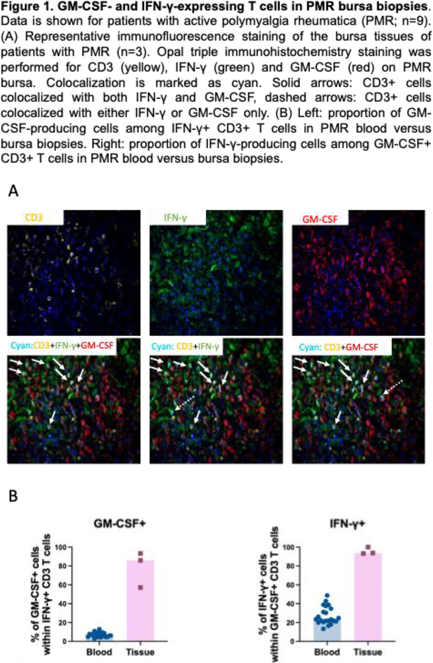
Results: The absolute numbers of CD3+, CD4+ and CD8+ T cells in peripheral blood were comparable between PMR and HCs. The percentages of CD4+ and CD8+ T cells expressing GM-CSF, IFN-γ, and IL-17 were similar in patients and controls. The majority of cytokine-producing CD4+ and CD8+ T cells expressed IFN-γ, GM-CSF, or both, with little IL-17 production. A substantial proportion of GM-CSF+CD4+ T cells co-produced IFN-γ. In contrast, only a small subset of GM-CSF+CD4+ T cells produced IL-17. Similar findings were observed for GM-CSF+CD8+ T cells. Immunohistochemistry on PMR biopsies showed moderate T cell infiltration, with most cells being CD4+ T cells. Opal triple immunofluorescence staining showed that GM-CSF and IFN-γ were abundantly expressed in the inflamed bursa tissue, and expression of these cytokines often co-localized with CD3 (Figure 1A). Moreover, GM-CSF and IFN-γ expression appeared more tightly linked in tissue than in peripheral blood, as evidenced by the high frequency of GM-CSF+CD3+ T cells co-expressing IFN-γ, and vice versa (Figure 1B).
Conclusions: In conclusion, the number of circulating GM-CSF-expressing T cells is unaltered in PMR. These cells predominantly co-express IFN-γ rather than IL-17. This association is even more pronounced in PMR tissue-infiltrating T cells where co-expression of IFN-γ and GM-CSF was even more common. Such dual cytokine-expressing T cells are well positioned to drive the differentiation of pro-inflammatory macrophages subsets previously described in PMR-affected tissues [1].
REFERENCES: [1] W. F. Jiemy et al. , “GM-CSF drives IL-6 production by macrophages in polymyalgia rheumatica,” (in eng), Ann Rheum Dis, vol. 84, no. 5, pp. 833-843, May 2025, doi: 10.1016/j.ard.2025.01.004.
[2] M. El-Behi et al. , “The encephalitogenicity of T(H)17 cells is dependent on IL-1- and IL-23-induced production of the cytokine GM-CSF,” (in eng), Nat Immunol, vol. 12, no. 6, pp. 568-75, Jun 2011, doi: 10.1038/ni.2031.
[3] G. Reynolds et al. , “Synovial CD4+ T-cell-derived GM-CSF supports the differentiation of an inflammatory dendritic cell population in rheumatoid arthritis,” (in eng), Ann Rheum Dis, vol. 75, no. 5, pp. 899-907, May 2016, doi: 10.1136/annrheumdis-2014-206578.
[4] J. Zhang et al. , “A novel subset of helper T cells promotes immune responses by secreting GM-CSF,” (in eng), Cell Death Differ, vol. 20, no. 12, pp. 1731-41, Dec 2013, doi: 10.1038/cdd.2013.130.
[5] R. D. Reitsema et al. , “Contribution of pathogenic T helper 1 and 17 cells to bursitis and tenosynovitis in polymyalgia rheumatica,” (in eng), Front Immunol, vol. 13, p. 943574, 2022, doi: 10.3389/fimmu.2022.943574.
Acknowledgments: NIL.
Disclosure of Interests: Anqi Zhang: None declared, Wayel Abdulahad: None declared, William F. Jiemy: None declared, Rosanne Reitsema: None declared, Yannick van Sleen: None declared, Maria Sandovici received consulting fees from Abbvie which were paid to the institution, Arjan Diepstra reports research support from Takeda, Thomas Kwee: None declared, Caroline Roozendaal: None declared, Peter Heeringa: None declared, Elisabeth Brouwer received a speaker fee for a talk on GCA at a post EULAR symposium in the Netherlands in 2023, Elisabeth Brouwer reports personal fees from Roche, outside the submitted work, Elisabeth Brouwer received grants from the Dutch Arthritis Society DAS and the EU/EFPIA/Innovative Medicines Initiative 2 Joint Undertaking Immune-Image which were paid to the Institution, Elisabeth Brouwer received compensation from Member Board nonprofit organization ARCH (Auto-immune Research Hub) in the Netherlands, which were paid to the institution in 2023, Kornelis van der Geest has received speaker fees from Roche and Orca Medical, unrelated to the submitted work, and an honorarium for editorial activities from Springer Nature, Kornelis van der Geest reports receiving research support from AbbVie and Siemens Healthineers.