fetching data ... 
Background: Structural damage progression is a major outcome in rheumatoid arthritis (RA). While its evaluation ideally involves radiographic scoring by two readers, this approach is challenging in large longitudinal cohorts. We previously demonstrated that a multireader assessment of radiographic structural damage progression is comparable to reference assessments and could improve the feasibility of radiographic scoring in large longitudinal cohorts followed over 5 years [1]. However, after 20 years, even using a multireader approach, the complete evaluation of an entire cohort remains a challenge. So, we built an algorithm, RADAR (Rheumatoid Arthritis Damage Automatic Reader) on a training part of a cohort of RA (BCD cohort) [2], we validated it on another part of this RA cohort (BCD cohort), and then we used it on an entire cohort of RA, the ESPOIR cohort [1].
Objectives: To demonstrate that an artificial intelligence algorithm enables the evaluation of an entire cohort’s radiographic data.
Methods: Studied population: The BCD cohort included 545 RA patients with radiographs of hands and feet at inclusion, 1 year, and 2 years, scored by four expert readers (two couples) using the modified van der Heijde Sharp score. The ESPOIR cohort included 813 patients between 2003 and 2005 with early arthritis (<6 months), a high probability of RA, and no prior DMARD treatment, who had radiographs of hands and feet every 6 months for 2 years, at years 2, 3, and 5, and then at years 10, 12, 15, and 20. They were previously read using modified van der Heijde Sharp Score [1]. In the ESPOIR cohort, during follow-up, 118 patients received another diagnosis (13.4%) and 110 died (13.5%). Algorithm: The Rheumatoid Arthritis Damage Automatic Reader (RADAR) algorithm was trained using 349 images, validated using 88 images and then tested on 108 images of the BCD cohort. RADAR detected radiographic characteristics (hand or foot, one or two sites per image, right or left), selected images without artifact that could interfere with reading (e.g., superimposition, poor orientation), and scores them using the modified van der Heijde Sharp score.
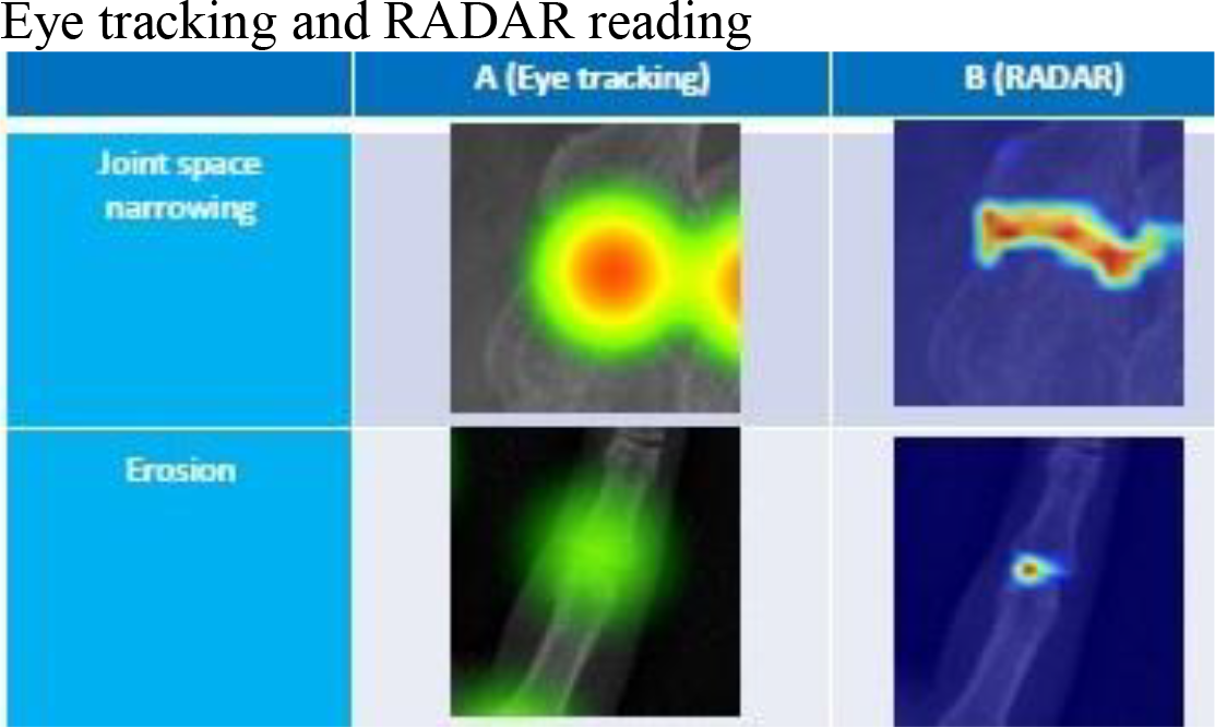
Comparison between human reading and artificial intelligence reading: We used human reading as a gold standard (mean of two readers) to evaluate sensitivity, specificity and accuracy of RADAR. We compared on selected X-Rays abnormalites detected by readers and artificial intelligence using eye-tracking (3) and Grad-CAM heatmaps, respectively.
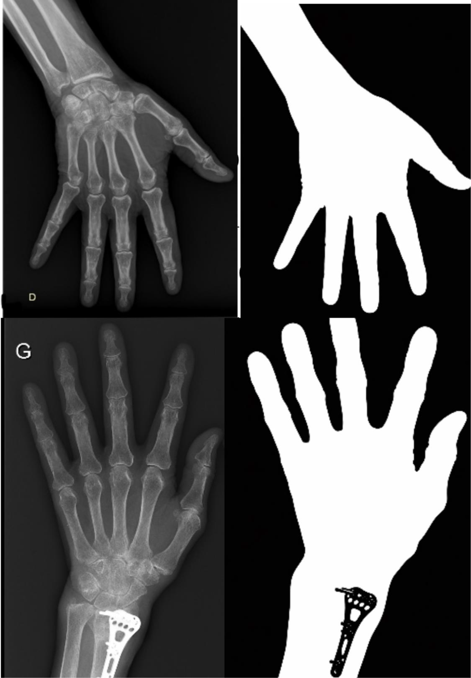
Results: RADAR was created and successfully analyzed a total of 7,560 joints from hand and foot radiographs on BCD cohort. These results remained consistent across the full spectrum of joint damage, from healthy joints to highly eroded ones. Reliability between the four human experts, evaluated on 30 radiographs, was good (κ = 0.7–0.9). Model performance metrics confirmed high agreement with expert readings: Binary classification: Accuracy = 0.94, Sensitivity = 0.92, Specificity = 0.95 (κ = 0.88). Multiclass (JSN) classification: Accuracy = 0.89, Sensitivity = 0.86, Specificity = 0.91 (ICC = 0.83). Grad-CAM heatmaps visually confirmed that the model focused on relevant joint regions, enhancing interpretability and clinical confidence. Figure 1 illustrates examples of joint detection by both human readers (eye tracking) and the RADAR pipeline. Then we read on the ESPOIR cohort which used heterogeneous imaging (jpeg for old x-rays which were scanned from argentic radiographs, DICOM for more recent images; in some cases only one hand by X-rays, in other two hands). First evaluations were performed on a curated subset of the espoir cohort including only radiographs with good quality. On this subset, binary classification achieved an estimated accuracy=0.95 sensitivity = 0.93, specificity=0.99 (ICC=0.86). Current efforts are now focused on preprocessing to read X-rays with artifacts in order to remove superfluous element of radiographs, reduce image variability and standardize the inputs provided by the models to improve robustness and generalizability (Figure 2).
Conclusions: RADAR demonstrates high accuracy and reliability in detecting and classifying joint damage in RA radiographs. It enables the evaluation of X-rays in a 20-year cohort of patients with early RA using artificial intelligence.
Eye tracking and RADAR reading
Preprocessing to read images with artifacts
REFERENCES: [1] Gandjbakhch F et al. Multireader assessment as an alternative to reference assessment to improve the detection of radiographic progression in a large longitudinal cohort of rheumatoid arthritis (ESPOIR). RMD Open. 2017 Jan 4;3(1):e00034.
[2] Garmendia A et al. Etude BCD. Revue du rhumatisme. 2025. A12-A21.
[3] Quere B et al. Can eye-tracking help to create a new method for X-ray analysis of rheumatoid arthritis patients, including joint segmentation and scoring methods. 2024; 7;3:e0000616.
Acknowledgments: NIL.
Disclosure of Interests: None declared.