fetching data ... 
Background: Spinal structural progression in axial spondyloarthritis is classically associated with inflammation, particularly inflammatory corner lesions. However, intervertebral ossification follows a variable and still incompletely understood process, involving factors other than inflammation, which may explain the presence of structural progressors without local inflammation and vice versa.
Objectives: The aim of this study was to demonstrate, using X-ray phase-contrast imaging, that ossification occurs upstream of both systemic and local inflammation in a model of reactive arthritis, adjuvant-induced arthritis (AIA).
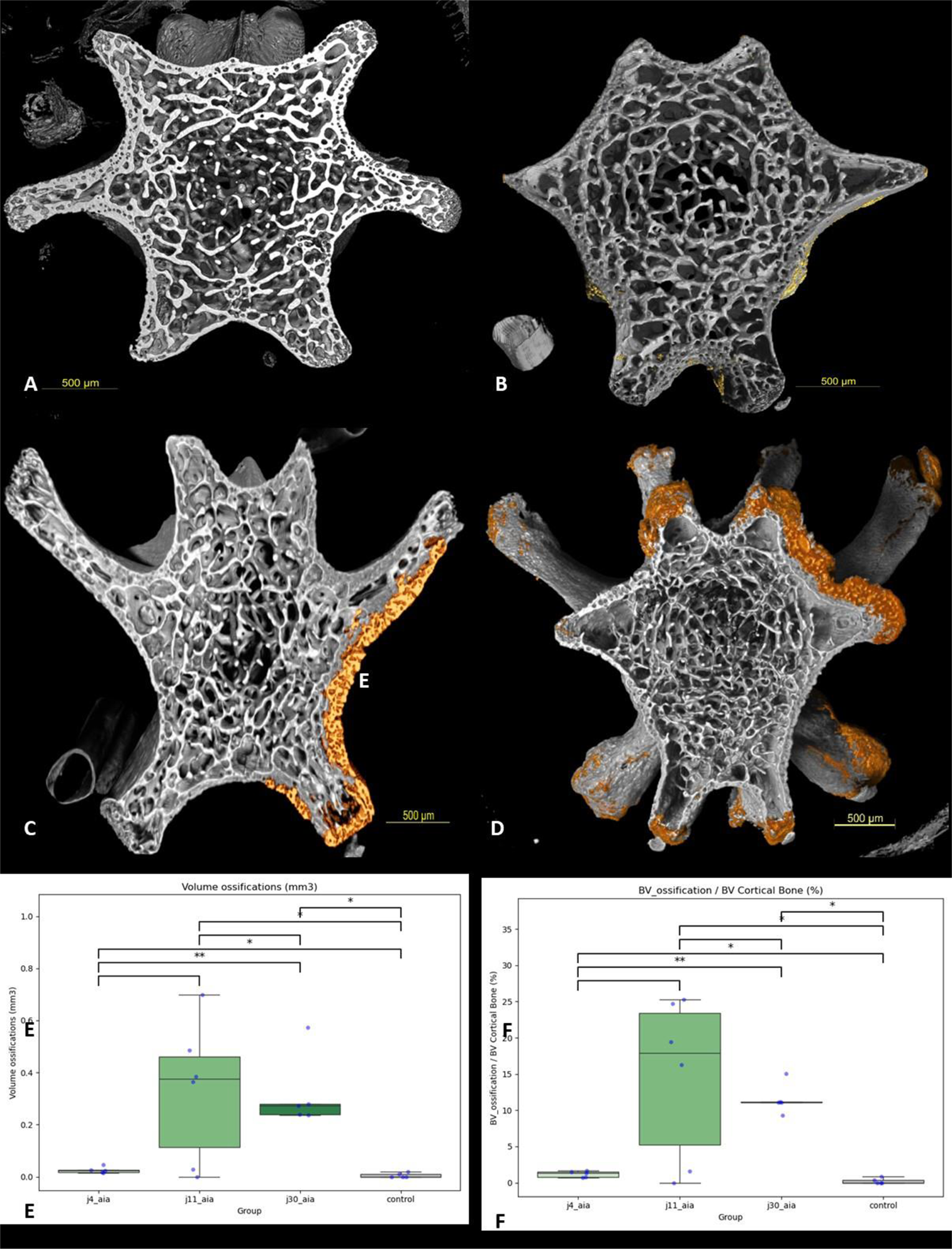
Methods: Arthritis was induced in 6-week-old male Lewis rats by subcutaneous injection of Mycobacterium butyricum . Analyses were performed in control rats and at three key stages of peripheral arthritis development in AIA rats: preclinical phase (day 4 post-induction), onset phase (day 11), and acute phase (day 30). A 2-cm tissue sample from the proximal tail and a terminal blood collection were performed. X-ray phase-contrast tomography of the tail sample was performed at the European Synchrotron Radiation Facility (ESRF, Grenoble). For each time point, automated analyses assessed bone mineral density, microarchitecture (bone volume, connectivity, degree of anisotropy, trabecular thickness), new bone formation, as well as the presence of tenosynovitis and erosions. Plasma levels of iFABP, P1NP, IL-1β, and TNF-α were measured by ELISA to evaluate systemic inflammation, bone formation, and intestinal barrier disruption.
Results: A total of five rats per group were included. A decrease in bone mineral density was observed as early as day 11, concomitant with increased TNF-α and IL-1β levels. Microarchitectural changes (Figure 1) were evident at day 11, including a significant decrease in trabecular thickness and connectivity (p<0.01). New bone formation was detected as early as day 4 at the periosteal surface of the metaphysis, at the joint capsule insertion (p<0.001), in parallel with increased levels of bone formation markers (P1NP; p<0.05) and intestinal barrier damage markers (iFABP; p<0.05). In addition, endosteal erosions were already present at day 4. Tenosynovitis was observed from day 11 onwards.
Conclusions: This study demonstrates that, in a model of reactive arthritis, early bone alterations and initiation of intervertebral ossification occur as early as day 4, preceding both systemic and local inflammation, in parallel with intestinal barrier disruption.
Subligamentous and perisoteal ossification over the time: Subligamentous and perisoteal ossification (in orange) in controls (A) and 4 days (B), 11 days (C) and 30 days (D) after the induction of the arthritis and comparison of the volume of the ossification in the different time point (E, F); * p<0.05; ** p< 0.01
REFERENCES: NIL.
Acknowledgments: NIL.
Disclosure of Interests: None declared.