fetching data ... 
Background: Osteoarthritis (OA) is a prevalent and painful disease that can affect multiple joints with the knee being the most commonly reported symptomatic site. While there are local anatomical changes within the knee joint, such as cartilage degeneration and synovitis that contribute to pain, it is well established that nervous system dysfunction also plays a critical role. Both peripheral sensitization of knee-innervating primary afferents and central changes within the spinal cord drive the persistence and chronicity of OA pain. Previous work from our laboratory demonstrated that prolonged oral administration of Δ 9 -tetrahydrocannabinol (THC) attenuated pain-related behaviours and disease progression in two mouse models of knee OA, including the destabilization of the medial meniscus (DMM) and monosodium iodoacetate (MIA) injection models. This analgesic effect was accompanied by transcriptional changes in distinct clusters of dorsal root ganglion (DRG) neurons, most notably Trpm8 + neurons. TRPM8 is a cold-sensitive ion channel previously implicated in analgesic mechanisms.
Objectives: This project seeks to investigate nervous system mechanisms underlying THC-mediated attenuation of osteoarthritis pain, with a focus on the potential contribution of TRPM8 + neurons.
Methods: Mice subjected to MIA (0.625 mg)-induced osteoarthritis received oral THC (10 mg/kg) or vehicle (medium-chain triglyceride, MCT) 5 days/week for 2 weeks and were sacrificed at 3 weeks post MIA injection. Ipsilateral L3-L5 dorsal root ganglia (DRGs) were collected, and single-nucleus RNA sequencing (snRNA-seq) was performed to assess THC-associated transcriptomic changes across distinct cell populations. Given that central sensitization and spinal hyperexcitability are key features of OA induced chronic pain, lumbar spinal cords were isolated in two independent experiments from saline- or MIA-injected mice, as well as from MIA-injected mice treated with MCT or THC. Immunofluorescence (IF) was used to quantify and spatially characterize neuronal activation based on co-expression of cFos and NeuN. To explore the potential contribution of TRPM8 + neurons to THC-associated modulation of osteoarthritis pain, a GFP-labelled Trpm8 DTR mouse line is being used whereby diphtheria toxin (DT; 100 ng) or saline (control) is administered intraperitoneally to selectively ablate TRPM8 + cells. Building on our initial IF characterization, spinal neuronal activation will be quantified following TRPM8 + cell ablation to assess deviations from baseline activation patterns. Ongoing studies will assess whether TRPM8 + cell ablation alters mechanical and thermal sensory behaviours associated with osteoarthritis and THC analgesia. In parallel, IF-based analyses will examine TRPM8 + neuronal distribution and innervation patterns in knee joints, L3-L5 DRG, and lumbar spinal cord to assess potential plasticity across OA progression and THC exposure.
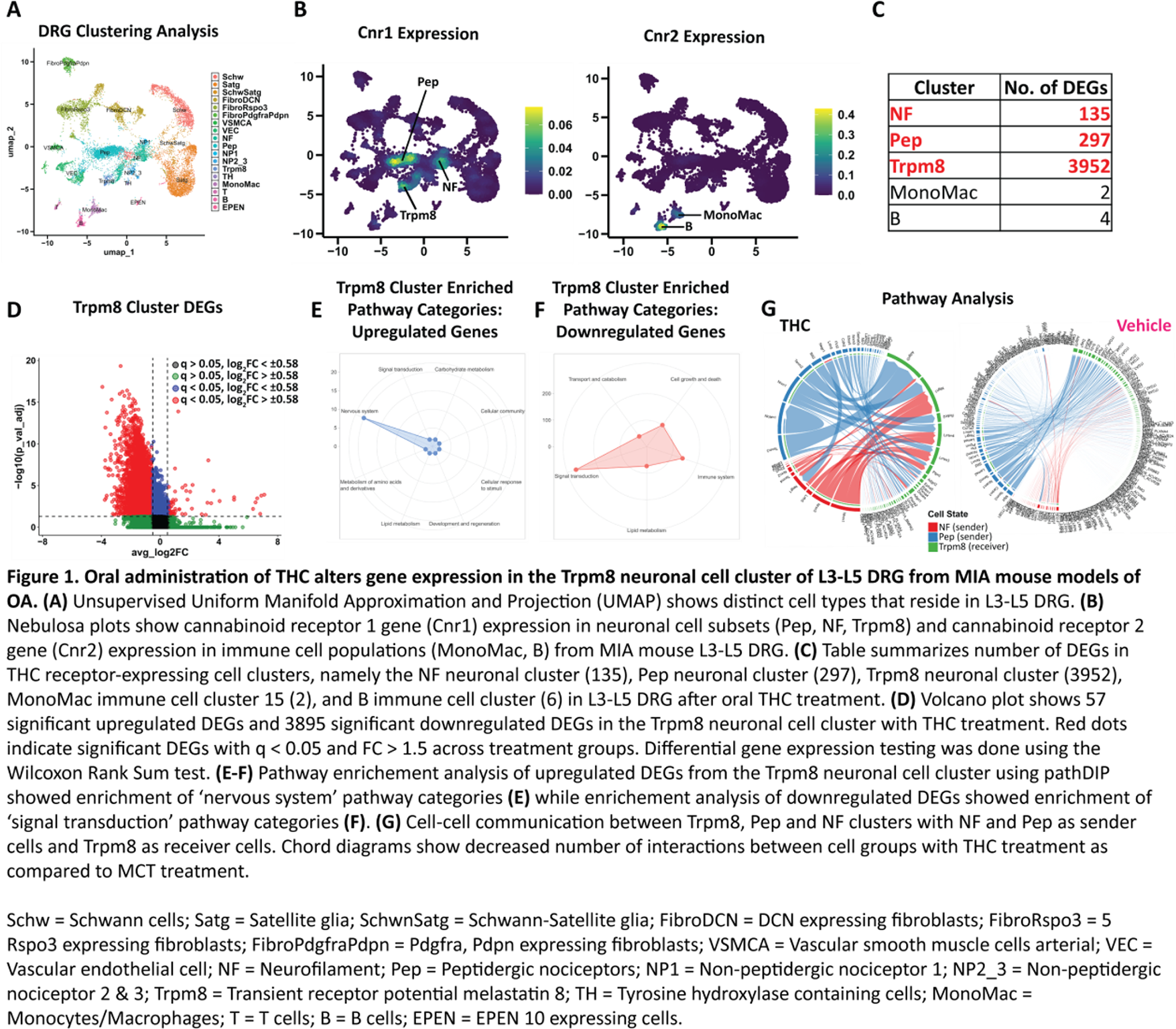
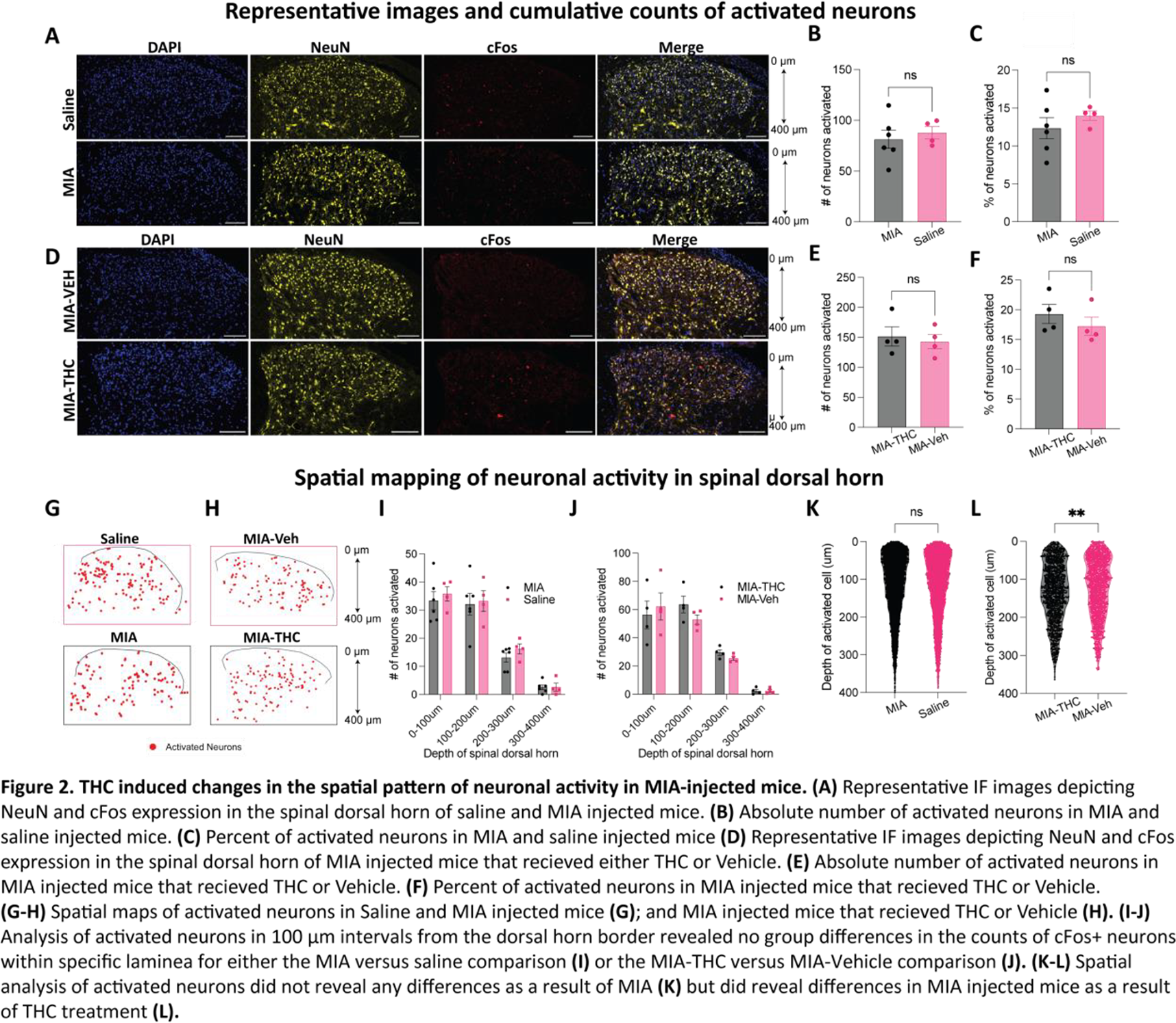
Results: SnRNA-seq analyses identified diverse cellular populations in joint-innervating L3–L5 dorsal root ganglia from MIA mice based on canonical marker gene expression ( Figure 1A , n=3/group). Cannabinoid receptor 1 gene ( Cnr1 ) and Cannabinoid receptor 2 gene ( Cnr2 ), the primary molecular targets of THC, were found in multiple neuronal subgroups including neurons positive for Trpm8 ( Figure 1B ). Among neuronal clusters, Trpm8 + neurons exhibited the largest number of differentially expressed genes following oral THC treatment, with 57 genes upregulated and 3,895 genes downregulated ( Figure 1C, D ). Pathway analysis of upregulated ( Figure 1E ) and downregulated ( Figure 1F ) genes within the Trpm8 neuronal cluster identified enrichment of nervous system-related pathways and signal transduction pathways, respectively, suggesting that THC treatment markedly impacts cellular pathways that shape neuronal signaling. Cell communication analyses suggested reduced ligand–receptor signalling between Trpm8 + neurons (receivers) and peptidergic/nociceptive neuronal populations (senders) following THC administration ( Figure 1G ). Preliminary IF analyses revealed no significant differences in either the proportion or absolute number of cFos + neurons across superficial or mid-to-deep regions of the spinal dorsal horn between MIA- and saline-injected mice ( Figure 2A-C ; n=4–6/group) or THC- and Vehicle-treated mice that had MIA injections ( Figure 2D-F ; n=4/group). Higher overall neuronal activation was observed within the superficial 200 µm of the dorsal horn, corresponding to laminae that receive TRPM8 + primary afferent input, as well as general nociceptive input ( Figure 2I, J ). However, this pattern did not differ between MIA- and saline-injected mice ( Figure 2G, I ) or between THC- and vehicle-treated MIA mice ( Figure 2H, J ). Spatial mapping of activated neurons ( Figure 2G, H ) indicated differences in the spatial distribution of cFos + neurons between THC- and vehicle-treated MIA mice ( Figure 2L ). In contrast, no differences in spatial activation patterns were observed between MIA- and saline-injected mice ( Figure 2K ). Together, these findings suggest that THC treatment is associated with altered spatial patterns of spinal neuronal activation in the absence of gross changes in overall activation levels.
Conclusions: Previous work from our group demonstrated that oral THC reduced pain-related behaviours in both DMM and MIA mouse models of KOA. In MIA mice, oral THC administration was associated with broad transcriptomic changes within Trpm8 + DRG neurons, highlighting this neuronal population as a potential target of cannabinoid action. Preliminary IF analyses further indicate THC-dependent alterations in the spatial distribution of neuronal activity within spinal cord regions innervated by TRPM8.
REFERENCES: NIL.
Acknowledgments: NIL.
Disclosure of Interests: None declared.