fetching data ... 
Background: Gastrointestinal (GI) manifestations are frequent in systemic sclerosis (SSc) and small-bowel involvement can lead to severe GI complications and malnutrition, contributing significantly to morbidity and mortality. A detailed understanding of molecular alterations of the small intestine is currently lacking but is critical for elucidating the underlying pathophysiology. T cells are of particular interest in gut health, since they are key regulators of mucosal immunity, support epithelial barrier integrity, and shape inflammatory responses.
Objectives: To characterize T cells and their interactions with the intestinal epithelium of the duodenum in SSc patients using high-definition spatial transcriptomics.
Methods: We evaluated duodenal biopsies from SSc patients collected in a standardized way in the randomized clinical ReSScue trial and healthy controls (HC). All SSc patients presented moderate to severe lower GI symptoms (UCLA SCTC GIT 2.0 score). Formalin-fixed paraffin-embedded (FFPE) samples were processed for immunohistochemistry (IHC), using anti-CD3, anti-CD4 and anti-CD8. QuPath software was used to determine the percentage of positively stained cells. Statistical analyses were performed using the Mann-Whitney test. FFPE samples were processed for spatial transcriptomics using the Visium HD Spatial Gene Expression Reagent kit v1. Libraries were sequenced on the Illumina NovaSeq X platform and aligned with Space Ranger v4. Data were analyzed using Seurat v5 and Banksy . We applied robust cell type deconvolution (RCTD), using a public scRNA-seq reference [1] to annotate cell types. Differentially expressed genes (DEG) were identified between SSc and HC using MAST . ClusterProfiler package was used for gene set enrichment analysis (GSEA). Module scores were calculated using the genes enriched in the significantly deregulated pathways. CellChat v2 was used for cell-cell interaction analysis.
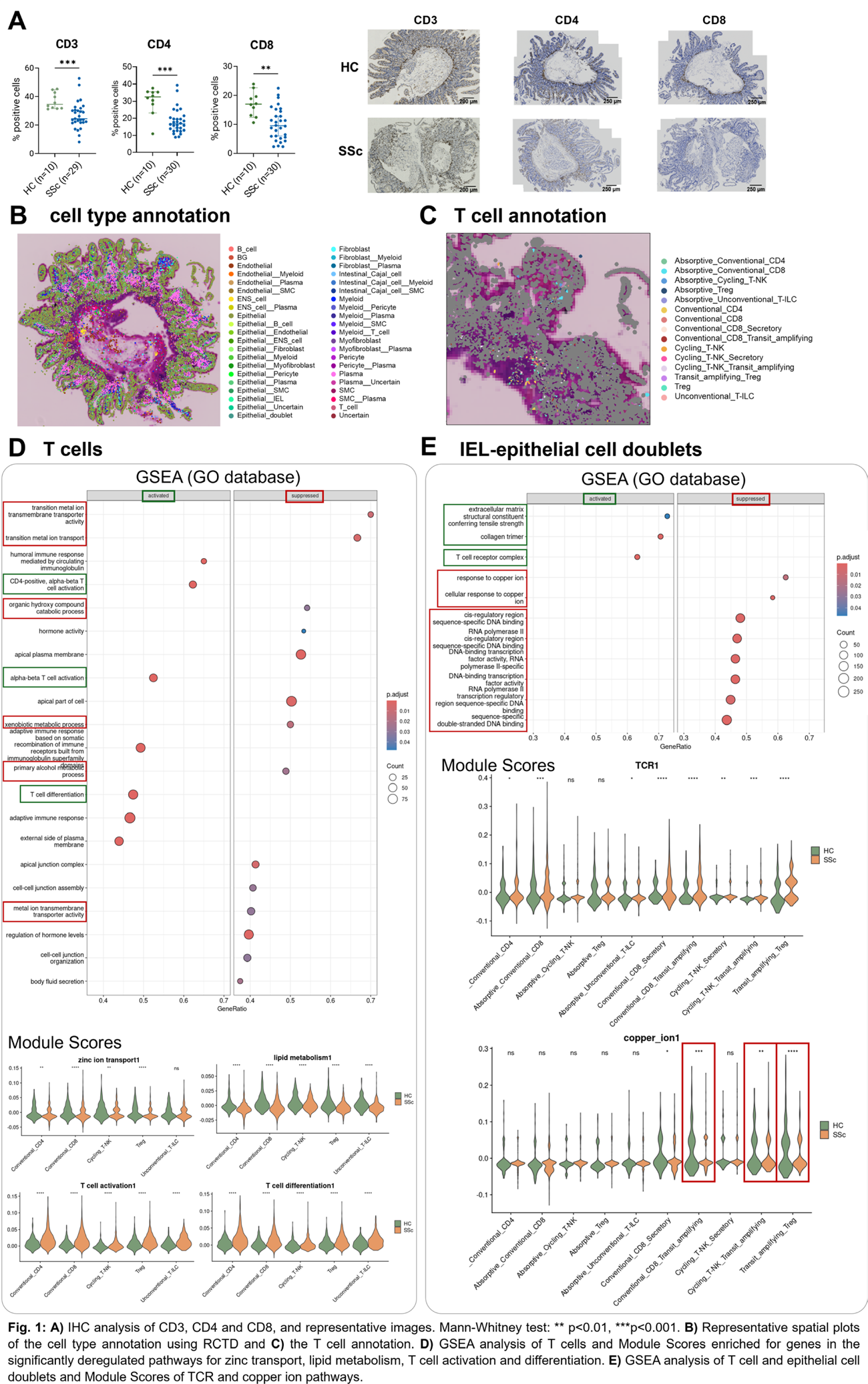
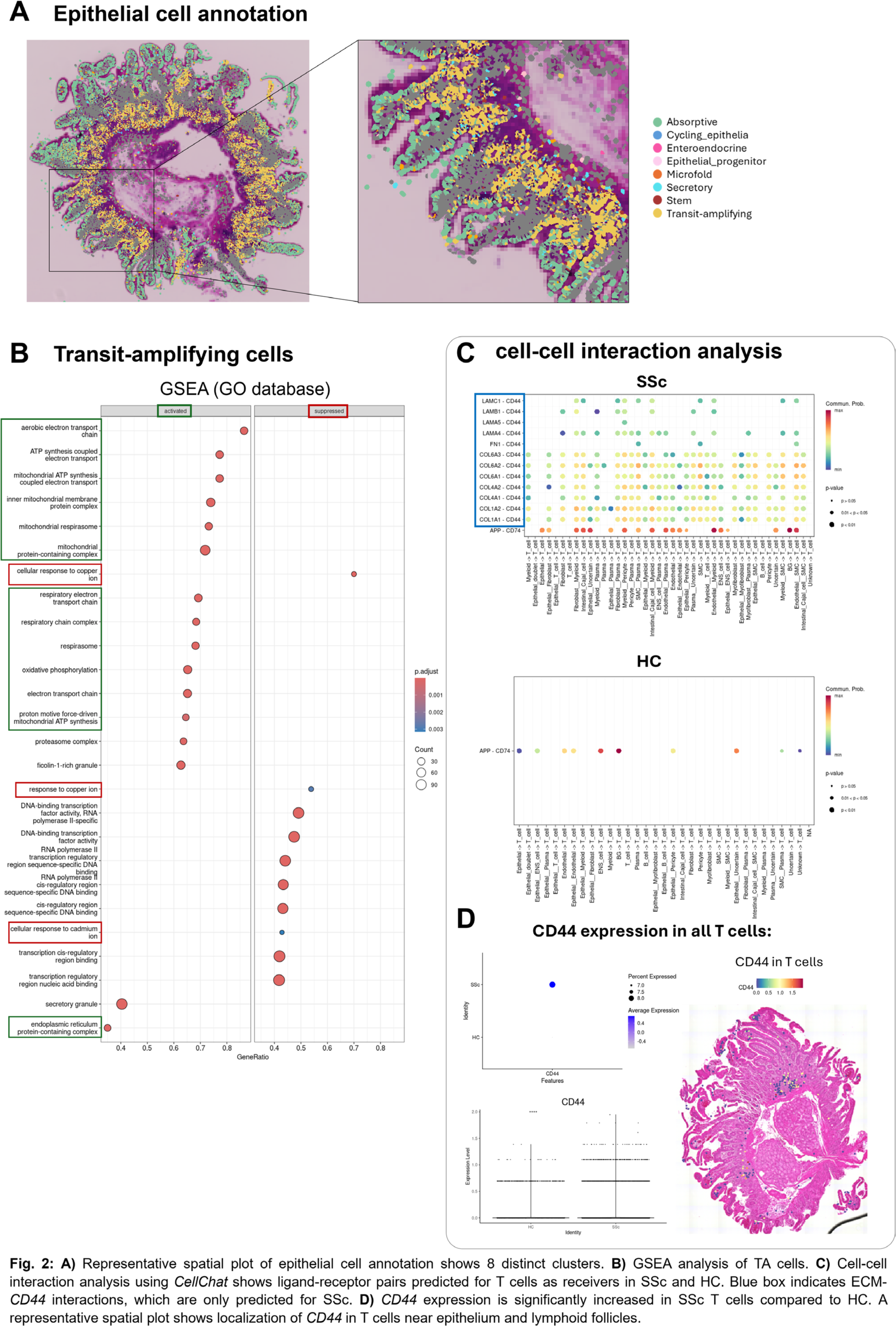
Results: IHC analysis revealed significantly reduced CD3 staining in SSc samples (n=29) compared with HCs (n=10). CD4 + and CD8 + T cells were also found to be significantly decreased in SSc compared to the HCs (n=10; Figure 1A). Together, these results indicate a reduced abundance of T cells in the SSc duodenum. Visium HD was performed on 19 SSc samples and 7 HCs and identified 42 distinct cell clusters (Figure 1B). T cell singlets were subsetted for downstream analyses (CD4 + , CD8 + , T regs , cycling T/NK and unconventional T cells; Figure 1C). GSEA identified reduced zinc transport and alcohol/lipid metabolism programs. CD8 + T cells and T regs showed the strongest decrease in zinc transport signatures as confirmed by gene module scores. Alcohol and lipid metabolism gene sets were suppressed in all T cell subsets. In contrast, pathways involved in T cell activation and differentiation were upregulated across all T cell subsets (Figure 1D). Our data suggest that, in the SSc duodenum, T cells are activated and recruited but are metabolically compromised, potentially leading to T cell exhaustion or dysfunction. Next, we analyzed intraepithelial lymphocyte (IEL) doublets with epithelial cells. We identified 10 distinct populations involving all T cell types (Figure 1C). GSEA showed upregulated TCR signaling in SSc. Module scores confirmed increased TCR activation in most IEL-epithelial doublets in SSc compared to HCs (Figure 1E). In addition, extracellular matrix (ECM) pathways were activated in SSc compared to HCs, likely reflecting an increased production of basement membrane collagens by epithelial cells. In contrast, copper-ion response pathways showed the strongest downregulation in doublets involving transit-amplifying cells with T regs , CD8 + T cells, or cycling T-NK cells. Pathways related to DNA binding and transcription factor (TF) activity were also reduced, with most of the enriched genes being general TFs abundant in T cells and epithelial cells (Figure 1E). These findings suggest that IELs in SSc become locally activated and may shift from a epithelial-supportive state toward a phenotype that potentially disrupts epithelial biology, particularly affecting transit-amplifying cells. We further characterized transit-amplifying cells, which are rapidly proliferating progenitor cells in the intestinal crypts (Figure 2A). In SSc, transit-amplifying cells showed upregulated mitochondrial pathways (e.g. “oxidative phosphorylation”, “electron transport chain”). Downregulated pathways were associated with zinc, copper, and cadmium responses, as well as DNA-binding and TF activity. Suppressed TFs included several regulators of cell cycle and proliferation (Figure 2B). Together, these data suggest that SSc transit-amplifying cells are less proliferative and potentially more differentiated, consistent with a metabolic shift toward oxidative phosphorylation. Cell-cell communication to assess T cell interactions revealed extensive collagen, fibronectin, and laminin signaling networks in SSc. T cells received these ECM signals through the cell-surface glycoprotein CD44 , which was significantly increased in SSc T cells compared to HCs (Figure 2C/D). Furthermore, CD44 , which is known to be involved in cell adhesion, was spatially localized near the epithelium and lymphoid follicles (Figure 2D). These interactions could contribute to T cell retention near the epithelium, consistent with our previous finding that T cells could modulate transit-amplifying cell behavior.
Conclusions: Our high-definition spatial analysis of a large SSc cohort from a randomized trial reveals shifts in T cell activation and epithelial dynamics in the small intestine. T cells exhibit strong activation signatures but simultaneously show signs of metabolic constraint, exhaustion, or dysfunction. CD44 -ECM interaction appears to anchor T cells near the epithelium, where they show increased activation compared to HCs and might impact adjacent transit-amplifying cells, driving them into a stressed state and potentially affecting their proliferation and differentiation.
REFERENCES: [1] Elmentaite et al. Nature 597, 250–255 (2021).
Acknowledgments: NIL.
Disclosure of Interests: Laura Much: None declared, Elena Pachera: None declared, Håvard Fretheim: None declared, Knut EA Lundin: None declared, Astrid Hofman: None declared, Øyvind Midtvedt: None declared, Espen Bækkevold: None declared, Lars Aabakken: None declared, Lumeng Li: None declared, Philip Stauffer: None declared, Maryam Asadikorayem: None declared, Shihan Xu: None declared, Blaz Burja Ely Lilly, Michael Scharl: None declared, Carina Mihai Boehringer Ingelheim, Janssen, Medbase, MED Talks Switzerland, Mepha, MedTrix, Novartis, PlayToKnow, Boehringer Ingelheim, Janssen, Medbase, MED Talks Switzerland, Mepha, MedTrix, Novartis, PlayToKnow, Øyvind Molberg: None declared, Oliver Distler Boehringer Ingelheim, 4P-Pharma, Abbvie, Acepodia, Aera, AnaMar, Anaveon, Argenx, AstraZeneca, Avalyn, Boehringer Ingelheim, BMS, Calluna, Cantargia, CSL Behring, EMD Serono, Galderma, Galapagos, Gossamer, Hemetron, Innovaderm, Kali, Lilly, Mediar, MSD Merck, Nkarta, Novartis, Oorja Bio, Orion, Pliant, Prometheus, Quell, Scleroderma Research Foundation, Skyhawk, Tandem, Topadur, UCB and Umlaut.bio, Kymera, Mitsubishi Tanabe, UCB, Anna-Maria Hoffmann-Vold Boehringer Ingelheim, Janssen, Medscape, Merck Sharp & Dohme, Novartis, Roche, AbbVie, Avalyn, Astra Zeneca, Boehringer Ingelheim, Bristol Myers Squibb, Calluna Pharma, Genentech, Janssen, Medscape, Merck Sharp & Dohme, Pliant, Roche, Werfen, Astra Zeneca, Boehringer Ingelheim, Janssen.