fetching data ... 
Background: Systemic sclerosis (SSc) is a systemic autoimmune disease characterized by fibrosis and immune dysregulation affecting the skin and internal organs. While inflammatory infiltrates have been observed in the skin of SSc patients, their cellular composition, origins, and clinical relevance remain unclear. Here we employed imaging mass cytometry (IMC) to characterize the cellular composition of immune cell aggregates and their cellular interactions and evaluate their value as tissue biomarkers.
Objectives: We employed imaging mass cytometry (IMC) to characterize the cellular composition of immune cell aggregates and their cellular interactions in SSc skin to evaluate their value as tissue biomarkers and potential therapy targets.
Methods: Skin punch biopsies from SSc patients were formalin-fixed, paraffin-embedded, and sectioned. Sections of 118 patients with SSc were screened by hematoxylin and eosin (H&E) histological staining for the presence of leukocyte aggregates or tertiary lymphoid structures. Imaging Mass Cytometry (IMC) was performed on the Hyperion platform (Standard BioTools) to collect data on the spatial distribution of protein targets and co-localization of cell populations within the skin tissue in four representative samples containing immune cell aggregates, eleven samples negative for lymphoid infiltrates and three samples derived from normal healthy skin.
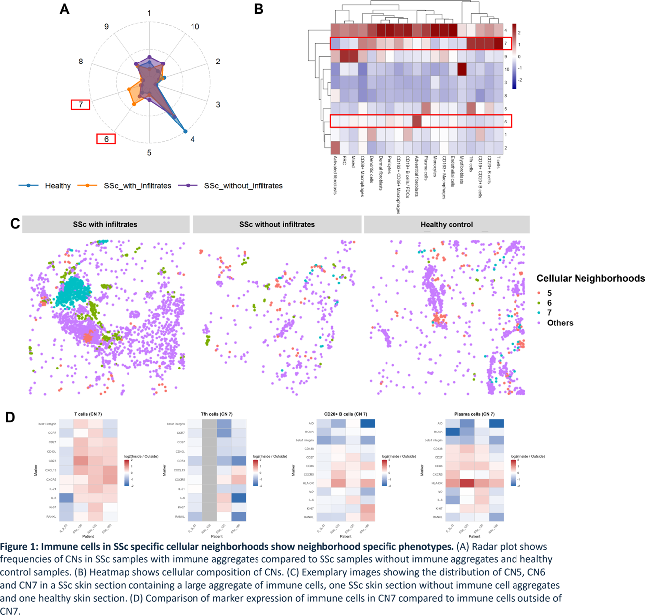
Results: Structured immune cell aggregates were identified in 14 of 118 H&E-stained SSc skin samples (12%). Large, diffuse lymphoid infiltrates were present in 19 samples (16%), whereas 13 samples (11%) showed no lymphoid infiltrates. Using our IMC panel, two distinct stromal–immune cellular neighborhoods corresponding to structured immune aggregates in SSc skin were identified. One neighborhood (CN7) was enriched for T follicular helper (Tfh) cells, CD20 + B cells, CD19 + CD20 + B cells, T cells, and plasma cells, whereas a second neighborhood (CN6) was characterized by adventitial fibroblasts expressing PDGFR β, PDGFR α, CXCL13 and CD34. Both CN6 and CN7 were significantly increased in SSc skin sections containing immune cell aggregates compared with SSc sections lacking aggregates and with healthy control skin (Figure 1). In sections containing large immune aggregates, CN6 and CN7 were consistently observed in close spatial proximity. Immune cells within CN7 exhibited phenotypic differences compared with the same cell types located outside of CN7. T cells in CN7 showed higher expression of CD40L, CXCL13, CXCR5, IL21, and IL6 relative to T cells outside CN7. CD73 expression was also increased in CN7-associated T cells. Although Tfh cells were infrequent overall, the majority were localized within CN7. Tfh cells in CN7 demonstrated increased expression of CXCR5 and CXCL13 and reduced expression of CD73 compared with Tfh cells outside CN7. Plasma cells within CN7 exhibited increased expression of CD138, CD27, CD86, CXCR5, and HLA-DR, along with reduced expression of BCMA, relative to plasma cells outside CN7. CD20 + B cells in CN7 displayed a similar activation-associated expression profile when compared with CD20 + B cells located outside CN7.
Conclusions: Using IMC, we provide first data on the cellular composition, the topographical organization and the cellular interactions within immune cell aggregates in SSc, as well niche-specific phenotypical changes induced in aggregating immune cells. T cells and Tfh cells located within immune cell aggregates showed upregulation of follicular markers such as CD40L, CXCL13, CXCR5, IL21, and IL6. These phenotypical changes indicate functional changes of those cells located inside the immune cell aggregates. T cells and Tfh cells shift towards B cell supporting activity by upregulation of follicular markers, in particular CD40L and IL-6. B cells and plasma cells showed high levels of activation and a reduced dependency on BCMA derived survival signals. These data indicate a possible formation of tertiary lymphoid structures in SSc skin, as CXCL13+ T cells have been described to promote formation of tertiary lymphoid structures (TLS) in cancer and other markers of TLS such as CXCL5, IL21, IL6, CD20, CD138 and CD3 are elevated within the described immune cell aggregates. Targeted inhibition of the formation of these structures and thereby interference with tissue resident immune responses in SSc could result in novel treatment options for SSc.
REFERENCES: NIL.
Acknowledgments: NIL.
Disclosure of Interests: None declared.