fetching data ... 
Background: Scleroderma renal crisis (SRC) is a life-threatening complication of systemic sclerosis (SSc). It presents with acute hypertension and rapid renal function decline, with 50% of cases manifesting thrombotic microangiopathy (TMA). Despite prompt angiotensin-converting enzyme inhibition and glucocorticoid avoidance, progression to the end-stage kidney disease and mortality remains frequent.
Objectives: This study aimed to map the cellular and molecular landscape of SRC kidneys via spatially-resolved, multiplex omics.
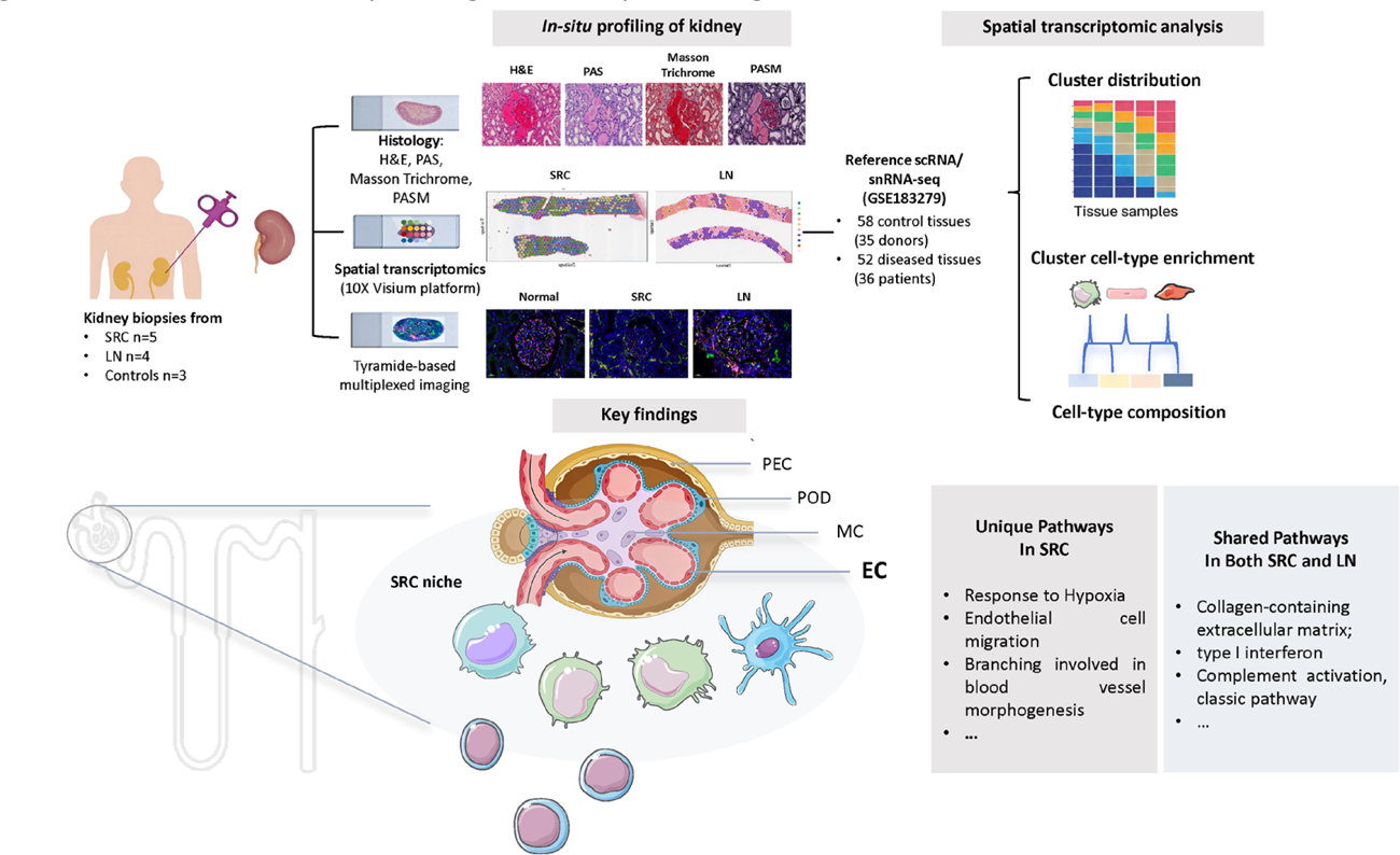
Methods: SRC kidney biopsies (n=5) were collected and compared with lupus nephritis (LN) (n=4) and histologically normal kidney tissue (n=3) obtained from non-malignant regions adjacent to benign renal tumors. Each sample underwent pathological review, followed by spatial transcriptomics profiling using the 10x Visium platform. Cell2location was used to deconvolute cell-type mixtures per Visium spot with the reference dataset of single-cell and single-nucleus RNA-sequencing, including 58 control tissues (35 donors) and 52 diseased tissues (36 patients). CellCharter was conducted to define, characterize and compare disease-specific cellular niches. Key findings were further validated using a 15-plex tyramide signal amplification (TSA)-based multiplex imaging assay.
Results: The 27 characterized renal and immune cell populations were resolved on kidney biopsies. SRC kidneys displayed fibroblast expansion and scattered immune infiltration, accompanied by the typical histological feature of TMA. Principal Component Analysis (PCA) exhibited a distinct cellular signature based on the cell composition within the compartment across the three different groups. Differentially expressed gene (DEG) analysis revealed 751 shared upregulated genes in both SRC and LN, which can be overrepresented in shared pathways mostly related to complement activation, type Ⅰ interferon production, and matrix biology. Moreover, SRC showed a unique signature, such as response to hypoxia and endothelial migration, whereas LN was enriched for the signaling of B cell activation and differentiation. CellCharter algorithm revealed SRC-specific cellular niches comprising resident renal cells (endothelial cells, podocytes, and parietal epithelial cells) and myeloid immune cells, while the LN microenvironment was more complexed with T cells, B cells, dendritic cells, and macrophages. Across SRC-specific niches, complement ( C1QA ), extracellular-matrix components ( COL1A1 , COL3A1 ), and key fibrosis regulators ( TGFBI , PDGFRB , ZEB2 ) were remarkably up-regulated. Consistently, overall kidney-level expression revealed even more pronounced complement activation, with C3 and C1QA/B/C all significantly increased, potentially shaping the pathological landscape of SRC. Spatial transcriptomics and TSA multiplex staining were employed to confirm the co-localization of C3 + and C1Q + cells within the vasculature of SRC kidneys.
Conclusions: These results highlight a spatially conserved and distinct injury program in SRC, which may inform future treatment approaches by targeting complement activation and fibrotic remodeling.
Overview of study design and key findings
REFERENCES: [1] Lake BB, Menon R, Winfree S, Hu Q, Melo Ferreira R, Kalhor K, Barwinska D, Otto EA, Ferkowicz M, Diep D, Plongthongkum N, Knoten A, Urata S, Mariani LH, Naik AS, Eddy S, Zhang B, Wu Y, Salamon D, Williams JC, Wang X, Balderrama KS, Hoover PJ, Murray E, Marshall JL, Noel T, Vijayan A, Hartman A, Chen F, Waikar SS, Rosas SE, Wilson FP, Palevsky PM, Kiryluk K, Sedor JR, Toto RD, Parikh CR, Kim EH, Satija R, Greka A, Macosko EZ, Kharchenko PV, Gaut JP, Hodgin JB; KPMP Consortium; Eadon MT, Dagher PC, El-Achkar TM, Zhang K, Kretzler M, Jain S. An atlas of healthy and injured cell states and niches in the human kidney. Nature. 2023 Jul;619(7970):585-594. doi: 10.1038/s41586-023-05769-3. Epub 2023 Jul 19. PMID: 37468583; PMCID: PMC10356613.
[2] Woodworth TG, Suliman YA, Li W, Furst DE, Clements P. Scleroderma renal crisis and renal involvement in systemic sclerosis. Nat Rev Nephrol. 2018 Feb;14(2):137. doi: 10.1038/nrneph.2017.183. Epub 2018 Jan 2. Erratum for: Nat Rev Nephrol. 2016 Nov;12(11):678-691. doi: 10.1038/nrneph.2016.124. PMID: 29292373.
[3] Cole A, Ong VH, Denton CP. Renal Disease and Systemic Sclerosis: an Update on Scleroderma Renal Crisis. Clin Rev Allergy Immunol. 2023 Jun;64(3):378-391. doi: 10.1007/s12016-022-08945-x. Epub 2022 Jun 1. PMID: 35648373; PMCID: PMC10167155.
[4] Yamashita H, Kamei R, Kaneko H. Classifications of scleroderma renal crisis and reconsideration of its pathophysiology. Rheumatology (Oxford). 2019 Dec 1;58(12):2099-2106. doi: 10.1093/rheumatology/kez435. PMID: 31566243.
[5] Del Galdo F, Lescoat A, Conaghan PG, Bertoldo E, Čolić J, Santiago T, Suliman YA, Matucci-Cerinic M, Gabrielli A, Distler O, Hoffmann-Vold AM, Castellví I, Balbir-Gurman A, Vonk M, Ananyeva L, Rednic S, Tarasova A, Ostojic P, Boyadzhieva V, El Aoufy K, Farrington S, Galetti I, Denton CP, Kowal-Bielecka O, Mueller-Ladner U, Allanore Y. EULAR recommendations for the treatment of systemic sclerosis: 2023 update. Ann Rheum Dis. 2025 Jan;84(1):29-40. doi: 10.1136/ard-2024-226430. Epub 2025 Jan 2. PMID: 39874231.
[6] Kuppe C, Ibrahim MM, Kranz J, Zhang X, Ziegler S, Perales-Patón J, Jansen J, Reimer KC, Smith JR, Dobie R, Wilson-Kanamori JR, Halder M, Xu Y, Kabgani N, Kaesler N, Klaus M, Gernhold L, Puelles VG, Huber TB, Boor P, Menzel S, Hoogenboezem RM, Bindels EMJ, Steffens J, Floege J, Schneider RK, Saez-Rodriguez J, Henderson NC, Kramann R. Decoding myofibroblast origins in human kidney fibrosis. Nature. 2021 Jan;589(7841):281-286. doi: 10.1038/s41586-020-2941-1. Epub 2020 Nov 11. PMID: 33176333; PMCID: PMC7611626.
Acknowledgments: NIL.
Disclosure of Interests: None declared.