fetching data ... 
Background: Immunogenetics strongly link psoriasis, psoriatic arthritis (PsA) and axial spondyloarthritis (axSpA) to IL-23/Th17 immune cell pathway, yet IL-23 blockade was inefficacious in ankylosing spondylitis (AS). Our prior work showed markedly higher inducible IL-23 protein production from spinal peri-entheseal bone (PEB) compared with peripheral knee joint PEB (Macleod, T ARD 2025). To further investigate site-specific regulation of immune responses, we applied a perturbomics framework to systematically assess how myeloid and lymphoid stimulation reshapes transcriptional programmes in the spine and knee using single-cell RNA sequencing.
Objectives: To characterize the immune cell composition of human spine knee and hip PEB and define how anatomical context impacts the IL-23/IL-17 cytokine axis at baseline and following innate and adaptive immune stimulation.
Methods: Spine, hip and knee PEB samples were obtained during orthopaedic surgery. Immune cells were isolated by mechanical digestion then characterized by multiparametric flow cytometry at baseline and following stimulation. In parallel, unsorted PEB immune cells were stimulated ex vivo with LPS, zymosan, IL-1β+anti-CD3 or anti-CD3 alone, and IL-23+anti-CD3. Cytokine production was quantified after 24 hrs (for LPS/zymosan stimulation) and 48 hrs (for CD3 stimulations) using ELISA and LEGENDplex assays. For transcriptional profiling, single-cell RNA-seq libraries were generated from approximately 10,000 unsorted immune cells per condition (unstimulated, LPS, anti-CD3, anti-CD3+IL-23) using 10x Genomics Next GEM 3′ reagents from age- and sex-matched knee and spine. Sequencing was performed at ~50,000 reads per cell
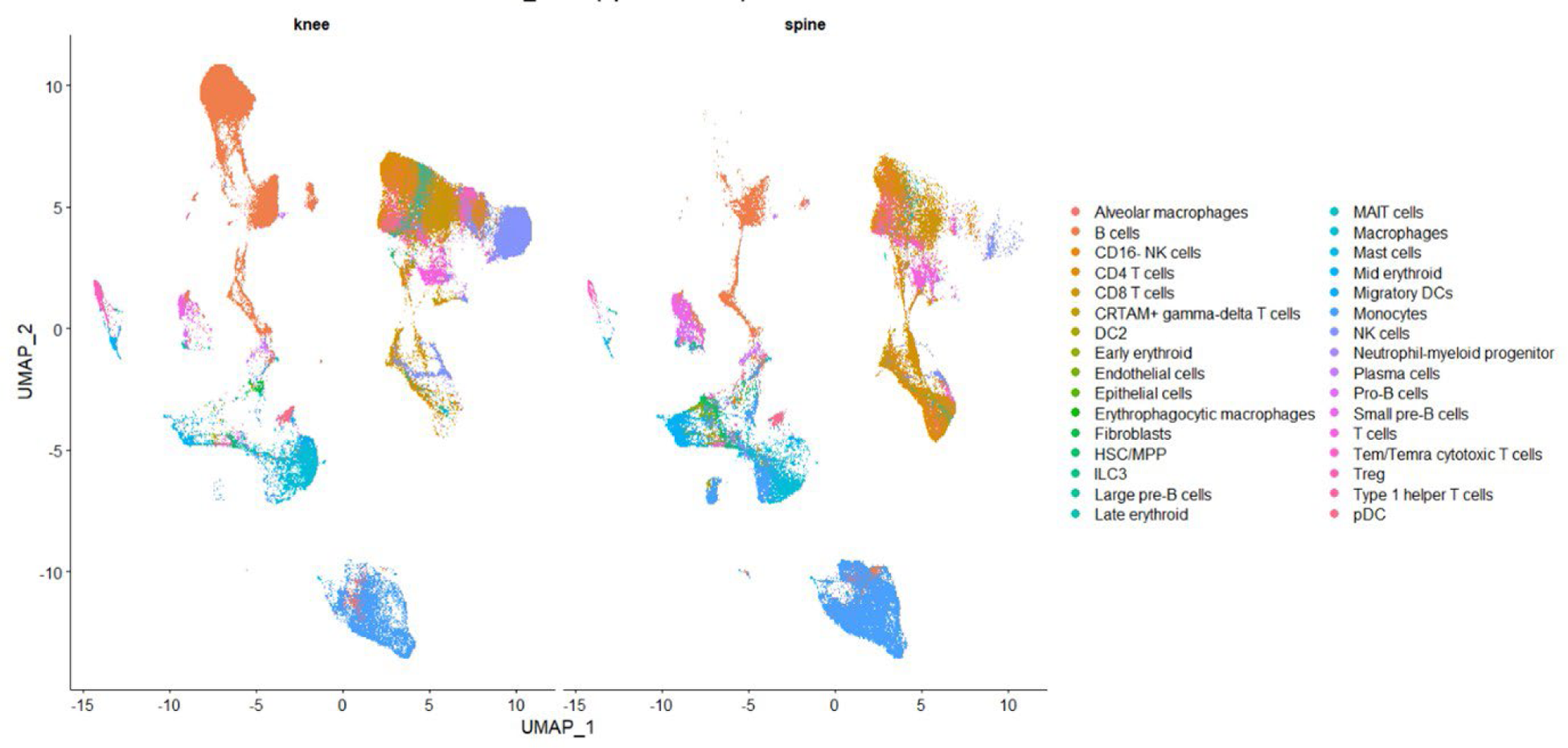
Results: Spinal PEB yielded approximately 10–100-fold more immune cells/gram of tissue than the knee. Despite this marked difference in cellularity, all major immune lineages were conserved across sites Figure (1). Analysis using matched cell numbers revealed differences in immune composition, with spine PEB relatively enriched for classical and intermediate monocytes, pDC-like cells, CD4 + helper T cells, Tregs and MAIT/type-17-like subsets, whereas knee PEB showed higher representation of cytotoxic CD8 + T cells, NK cells and APC-like populations. Stimulation of ex vivo PEB cells demonstrated higher IL-23 production from spine-derived immune cells compared with knee. Single-cell transcriptomic analysis also showed robust IL23A upregulation in spine intermediate monocytes and APC-like clusters paired with IL12B upregulation following LPS stimulation, while knee myeloid cells exhibited only modest transcriptional induction. Single-cell analysis of T cell transcriptomes revealed higher IL17A / IL17F expression and type-17 programme scores in knee-derived T cells compared with spine, particularly following anti-CD3+IL-23 stimulation. Spine T cells, despite residing in an IL-23-producing environment, showed attenuated type-17 transcriptional responses. Induction of innate inflammatory cytokines such as IL1B and TNF was observed predominantly within myeloid populations following LPS stimulation. Integrated module scoring demonstrated more efficient coupling between myeloid IL-23 production and T cell type-17 programme induction in knee samples, whereas spine samples exhibited relative uncoupling of the IL-23/type-17 axis across stimulation conditions.
Conclusions: These data demonstrate that while knee and spine PEB tissues share major immune cell lineages, they show differential IL-23 production capacity and IL-23–type 17 pathway coupling. Spine peri-entheseal bone exhibits a myeloid-rich, IL-23-producing environment with attenuated downstream T cell type-17 transcriptional responses, whereas knee tissue shows more responsiveness to IL-23 leading to type-17 programme induction. These site-specific differences in immune organisation provide further biological plausibility for understanding divergent immunological behaviours between axial and peripheral spondyloarthritis.
UMAP visualisation of single-cell RNA sequencing data from human knee (left) and spine (right) PEB. Each point represents a single cell, coloured by annotated immune and stromal cell type based on transcriptional profiling. Major immune lineages are present at both anatomical sites.
REFERENCES: [1] Macleod, T. et al. Annals of the Rheumatic Diseases, Volume 84, 508
Acknowledgments: NIL.
Disclosure of Interests: Tom Macleod This research project was funded by Johnson & Johnson., Mark Harland This research project was funded by Johnson & Johnson., Chi Wong This research project was funded by Johnson & Johnson., Kerem Abacar This research project was funded by Johnson & Johnson., Yaaseen Hamid This research project was funded by Johnson & Johnson., Abhay S Rao This research project was funded by Johnson & Johnson., Almas Khan This research project was funded by Johnson & Johnson., Peter Loughenbury This research project was funded by Johnson & Johnson., Vishal Borse This research project was funded by Johnson & Johnson., Cambell MacEachern This research project was funded by Johnson & Johnson., Antonius Pieter Van den Heuvel by Johnson & Johnson., Warner Chen by Johnson & Johnson., Ala Altaie This research project was funded by Johnson & Johnson., Dennis McGonagle This research project was funded by Johnson & Johnson.