fetching data ... 
Background: Systemic lupus erythematosus (SLE) is characterized by dysregulated cytokine and chemokine networks, with type I interferons (IFNs) as key pathogenic drivers; however, their extremely low serum levels limit accurate quantification by conventional ELISA. Moreover, because type I, II, and III IFNs share common signaling pathways, their type-specific functions in immune cells remain incompletely defined. Although proteomic studies have reported widespread cytokine and chemokine alterations in SLE, limited concordance across platforms such as Olink and SomaScan and strong correlations with IFN activity have hindered robust identification of IFN-independent immune functions. To address these challenges, we applied ultrasensitive digital ELISA for precise IFN quantification and integrated multiple proteomic platforms to establish a reliable cytokine/chemokine measurement panel, enabling elucidation of immune cell–specific cytokine and chemokine functions in SLE.
Objectives: To elucidate the immune cell–cytokine/chemokine dynamics in SLE based on the precise quantification of IFNs.
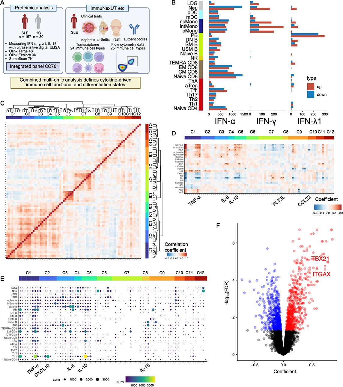
Methods: We performed an integrated analysis of transcriptomic, proteomic, and immunophenotypic data across up to 25 immune cell types obtained from up to 157 patients with SLE and 30 healthy controls (HC), leveraging our immune-cell transcriptome database, ImmuNexUT ( Cell . 2021), and other previous reports ( Cell . 2022, Sci Immunol . 2024). Using stored serum samples, we quantified IFN-α, IFN-γ, IFN-λ1 and interleukin-1β (IL-1β) by ultrasensitive digital ELISA. Cytokine/chemokine profiling using Olink Target 48, Olink Explore 3K, and SomaScan 7K identified highly concordant analytes across platforms, which were integrated into a unified panel, hereafter referred to as CC76. In addition, machine learning–based automated gating was applied to flow cytometry data to generate accurate and reproducible immunophenotypic data. ImmuNexUT, the CC76 panel, and the high-quality immunophenotyping data were integrated to delineate the immune cell–cytokine/chemokine dynamics in SLE (Figure 1A).
Results: Using ultrasensitive digital ELISA, serum IFN-α, IFN-γ, and IFN-λ1 levels were detectable above the assay sensitivity threshold in 83.3% (105/126), 100% (126/126), and 99.1% (106/107) of patients, respectively. Serum IFN-α levels were strongly correlated with IFN-γ and IFN-λ1 levels, as well as with the expression levels of interferon-stimulated genes (ISG) in immune cell types (e.g., classical monocytes, r = 0.89). DEG analysis revealed that IFN-α, IFN-γ, and IFN-λ1 affect distinct immune cell subsets (Figure 1B). Next, using the CC76 panel, we performed a comprehensive analysis of cytokine and chemokine expression. Compared with HC, patients with SLE in remission exhibited elevated levels of IFN-γ–related cytokines (IFN-γ and CXCL10), IL-10, IL-15, and CXCL13, defining distinct disease-state-associated cytokine/chemokine signatures. Hierarchical clustering of cytokine and chemokine expression measured by the CC76 panel grouped the analytes into 12 clusters (Figure 1C). After adjusting for IFN-driven effects, we evaluated the associations between cytokines/chemokines and clinical traits. IFN-λ1, IL-6, IL-10, and TNF-α were positively associated with SLEDAI-2K scores, proteinuria, and anti–dsDNA antibody titers. In contrast, FLT3LG and CCL22 were negatively associated with SLEDAI-2K and physician global assessment, respectively, suggesting potential immunoregulatory roles (Figure 1D). Further DEG analysis was performed after adjusting for IFN-driven effects (Figure 1E). Gene ontology analysis revealed IL-10 was associated with cell cycle pathways in Th1 cells, effector memory (EM) CD8 + T cells, and PBs. IL-6 and TNF-α were associated with cell cycle pathways in PBs and inflammatory pathways in classical monocytes. CXCL10 was also associated with cell cycle pathways in Th1 cells. IL-15 was associated with increased expression of ITGAX and TBX21, signature genes of double-negative 2 (DN2) B cells, in naïve B cells, suggesting a role for IL-15 in the differentiation of naïve B cells toward DN2 B cells (Figure 1F). These cytokine-driven transcriptional programs are likely to contribute to disease exacerbation in SLE.
Conclusions: By ultrasensitive quantification of type I, II, and III interferons and separation of their shared signaling effects, we defined interferon type–specific impacts on distinct immune cell subsets in SLE. Using a unified cytokine/chemokine panel (CC76), interferon-adjusted analyses linked cytokine- and chemokine–associated programs to activation of Th1 cells, effector memory CD8 + T cells, plasmablasts, and classical monocytes, as well as differentiation of naïve B cells toward DN2 B cells. These findings provide a refined framework for immune stratification in SLE and identify immune pathways beyond global interferon blockade as potential therapeutic targets. (A) Overview of this study. (B) Bar plots showing the number of DEGs associated with each IFN type. DEG analysis was performed using log-transformed serum concentrations of IFN-α, IFN-γ, and IFN-λ1, with sex and age included as covariates. (C) Heatmap of correlations among CC76 panel cytokines and chemokines, with hierarchical clustering based on expression levels. (D) Heatmap showing the effects of each cytokine/chemokine on clinical traits after adjustment for IFN-driven effects. Linear regression analyses were performed for each clinical trait with individual cytokines/chemokines as predictors and ISG expression, sex, and age included as covariates. (E) A bubble plot showing the number of DEGs associated with each cytokine/chemokine for each immune cell type. (F) A volcano plot of IL-15-associated DEGs in naïve B cells.
REFERENCES: NIL.
Acknowledgments: NIL.
Disclosure of Interests: Tatsuki Abe: None declared, Takahiro Itamiya T.I. belongs to the Social Cooperation Program, Department of functional genomics and immunological diseases, supported by Chugai Pharmaceutical., Yumi Tsuchida: None declared, Haruka Tsuchiya H.T. has received spearkers’ bereau from AbbVie, Amgen, Asahi Kasei, Astellas, Bristol-Myers Squibb, Chugai, Daiichi-Sankyo, Eisai, Eli Lilly, Gilead, Jansen, Novartis, Sanofi, Tanabe Mitsubishi and UCB, H.T. has received grant from AbbVie, Mochida, Takeda, Hirofumi Shoda: None declared, Mineto Ota M.O. has received speaker’s bureau honoraria from MSD, AstraZeneca, Chugai Pharmaceutical, and Calico Life Sciences., M.O. has received grants from GSK, the Mochida Memorial Foundation for Medical and Pharmaceutical Research, the Astellas Foundation for Research on Metabolic Disorders, and the Chugai Foundation for Innovative Drug Discovery Science. M.O. belonged to the Social Cooperation Program, Department of functional genomics and immunological diseases, supported by Chugai Pharmaceutical., Tomohisa Okamura T.O. belongs to the Social Cooperation Program, Department of functional genomics and immunological diseases, supported by Chugai Pharmaceutical., Keishi Fujio K.F. has received payments or honoraria from Chugai Pharmaceutical Co., Ltd,AbbVie GK,Asahi Kasei Pharma Co., Ltd, Bristol Myers Squibb K.K., AstraZeneca K.K., Mitsubishi Tanabe Pharma Corporation, Eisai Co., Ltd, Gilead Sciences K.K., Eli Lilly Japan K.K., Pfizer Japan Inc., Taisho Pharmaceutical Co., Ltd, Astellas Pharma Inc., Daiichi Sankyo Co., Ltd, Novartis Pharma K.K., GlaxoSmithKline K.K., and Alexion Pharma GK., K.F. has received research grants from Chugai Pharmaceutical Co., Ltd, AbbVie GK, Asahi Kasei Pharma Co., Ltd, Bristol Myers Squibb K.K., AstraZeneca K.K., Eisai Co., Ltd, Tsumura Co., Ltd, and Taisho Pharmaceutical Co., Ltd; consulting fees from Asahi Kasei Pharma Co., Ltd.