fetching data ... 
Background: Lupus nephritis (LN), a severe complication of systemic lupus erythematosus (SLE), affects 50%-70% of SLE patients, with 30%-40% developing treatment resistance and end-stage renal disease. Aberrant activation of innate immune cells (macrophages, neutrophils) and their infiltration into renal vasculature drive necrotizing arteritis and renal injury, but the upstream regulatory mediators remain unclear. CCL8, a C-C chemokine, mediates inflammatory cell recruitment via CCR2/CCR5, while MiT-TFE family transcription factors (MITF/TFEC) regulate macrophage activation in vascular diseases—yet their roles in LN remain unexplored. Umbilical cord blood-derived mesenchymal stem cells (UCB-MSCs) exhibit immunomodulatory potential, but their precise mechanism in LN, especially on macrophage-neutrophil crosstalk, is not fully elucidated.
Objectives: To systematically decipher the molecular mechanism by which UCB-MSCs alleviate LN in human patients and mouse models, focusing on the regulation of MiT-TFE/CCL8 signaling and CCL8 + macrophage-CCR5 + neutrophil crosstalk using single-nucleus RNA sequencing (snRNA-seq) and multi-omics validation.
Methods: Three complementary mouse models were used: MRL/lpr lupus-prone mice, CCL8 knockout (KO) mice with pristane-induced SLE, and R848-induced SLE mice. MRL/lpr mice (12-week-old, LN-established) were grouped into WT (C57BL/6J), LN (PBS), MSC-L (2×10 6 cells/mouse), and MSC-H (5×10 6 cells/mouse) with weekly tail vein injections for 4 weeks. Key techniques included snRNA-seq (10x Genomics, Illumina NovaSeq 6000) for renal cellular transcriptomics; network pharmacology (TCMSP, SwissTargetPrediction, OMIM) to predict target pathways; histological (H&E, IHC, IF), molecular (qPCR, Western blot, ELISA), and flow cytometry analyses for validation; Transwell assays for neutrophil chemotaxis. Core molecular signatures were verified in renal tissue samples from LN patients.
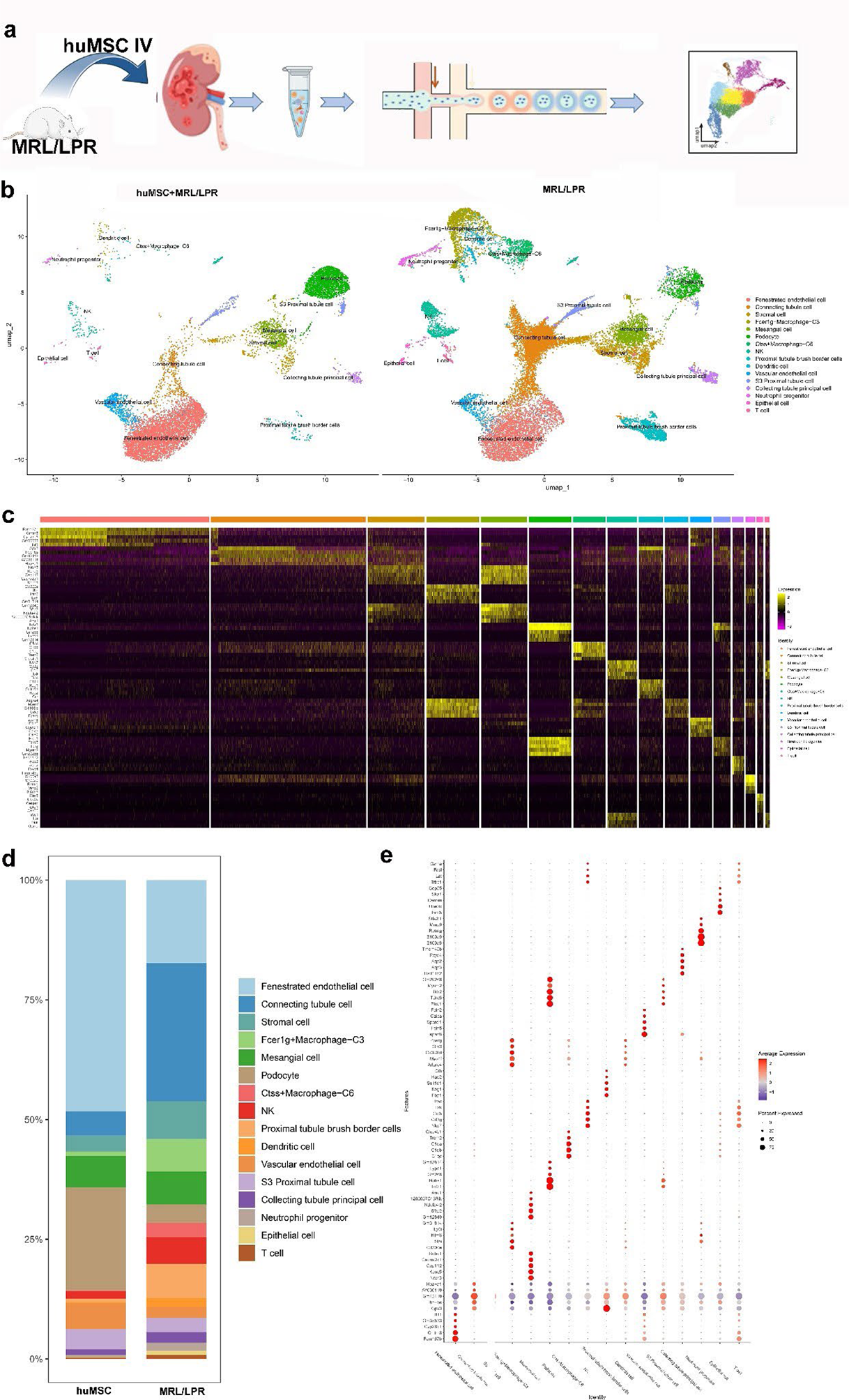
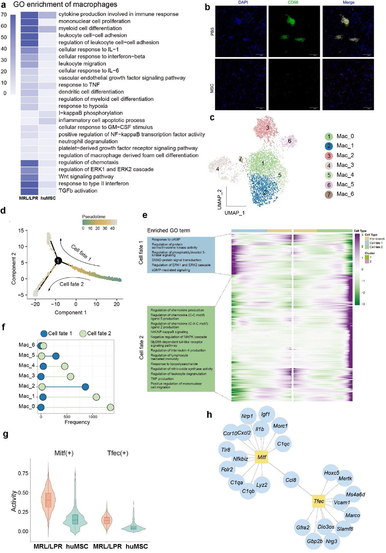
Results: UCB-MSCs exerted profound therapeutic effects across all SLE/LN mouse models. In MRL/lpr mice, UCB-MSCs (especially MSC-H) reduced serum anti-ds-DNA antibody levels (25-fold elevation in LN vs 6.3±1.1 U/mL in MSC-H, P<0.01), 24h urinary protein (385.6±42.3 vs 126.3±18.5 mg/24h, P<0.01), and serum creatinine (89.5±9.6 vs 58.7±7.1 μmol/L, P<0.05). Renal pathology scores (glomerular injury, interstitial inflammation, vasculitis) decreased from 8.7±1.2 (LN) to 3.2±0.8 (MSC-H, P<0.01), with mitigated myocarditis and abdominal aortic dilation, while CCL8 KO mice showed attenuated LN phenotypes with reduced neutrophil infiltration. SnRNA-seq captured 12,457-15,872 high-quality nuclei clustering into 15 cell types; LN kidneys had 3.2-4-fold higher macrophage proportion (12.7%±1.5% vs 3.2%±0.6% in WT, P<0.001) and 5-fold higher neutrophil proportion (8.9%±1.1% vs 1.8%±0.4%, P<0.001), which were restored by UCB-MSCs (macrophages: 5.8%±0.9%; neutrophils: 4.2%±0.7%), with DEGs in LN macrophages including upregulated MITF, TFEC, and CCL8 that were downregulated by UCB-MSCs. LN macrophages had 4-5-fold higher MITF/TFEC activity (P<0.001) with nuclear colocalization (co-localization coefficient 0.68±0.07); ChIP-qPCR confirmed MITF/TFEC directly bind the CCL8 promoter (enrichment folds: 3.8±0.4 and 3.2±0.3, P<0.01), and UCB-MSCs reduced MITF/TFEC activity by ~60% (P<0.01), decreasing CCL8 mRNA (5.2±0.6 vs 2.1±0.3-fold vs WT, P<0.01) and protein levels (IHC OD: 0.42±0.05 vs 0.23±0.03, P<0.01). CellphoneDB analysis showed CCL8-CCR2/CCR5 interaction scores increased 4.1-fold and 3.7-fold in LN (P<0.001), reduced to <30% by UCB-MSCs; flow cytometry revealed LN kidneys had 28.7% CCL8 + macrophages (F4/80 + CCL8 + ) and 31.2% CCR5 + neutrophils (MPO + CCR5 + ), dropping to 12.3% and 14.5% post-UCB-MSCs (P<0.05), with IF co-localization coefficient of CCL8 and CCR2/CCR5 decreasing from 0.72 to 0.29 (P<0.05). LN neutrophils had 35% Neu_1 subset (high CCR2/CCR5/PAD4) enriched in NETs formation; UCB-MSCs reduced Neu_1 proportion (42.5%±3.6% vs 23.8%±2.8%, P<0.01) and PAD4 expression (mRNA: 4.8±0.5 vs 2.0±0.3-fold, P<0.01), inhibiting NETs formation (Cit-H3 + /MPO + co-localization) and renal arteritis (MPO + infiltration score: 3.8±0.5 vs 1.7±0.3, P<0.01). Network pharmacology identified 48 core targets enriched in chemokine signaling, macrophage activation, and neutrophil chemotaxis pathways, with UCB-MSCs inhibiting p-ERK1/2 and p-NF-κB p65 in the chemokine pathway.
Conclusions: UCB-MSCs alleviate LN by targeting MiT-TFE family transcription factors (MITF/TFEC) to suppress CCL8 secretion from pro-inflammatory macrophages, thereby disrupting CCL8 + macrophage-CCR5 + neutrophil crosstalk and inhibiting neutrophil chemotaxis, NETs formation, and vasculitis. This mechanism is validated in multiple mouse models and human LN samples, providing a novel precision therapeutic strategy for LN by targeting innate immune cell interactions.
Single-nucleus transcriptomic remodeling of kidney tissue in MRL/LPR mice after human umbilical cord blood mesenchymal stem cell (huMSC) intervention
Functional enrichment of macrophage subsets and regulatory roles of MiT-TFE family transcription factors
REFERENCES: NIL.
Acknowledgments: NIL.
Disclosure of Interests: None declared.