fetching data ... 
Background: Accurate assessment of disease activity in axial spondyloarthritis (axSpA) is crucial for guiding therapeutic decisions, such as initiating or switching biologic DMARDs. However, determining the inflammatory status remains challenging due to the limited sensitivity of physical examinations and the subjective nature of patient-reported outcomes. While Magnetic Resonance Imaging (MRI) is widely regarded as a reliable modality for detecting active inflammation, visual interpretation is labor-intensive and subject to significant inter-observer variability. To date, no standardized, automated system exists to quantify global disease activity by synthesizing information from the entire axial skeleton. We hypothesized that an artificial intelligence model could objectively replicate a specialist’s comprehensive assessment.
Objectives: This study aimed to develop and validate a multi-modal deep learning model capable of predicting axSpA disease activity (active vs. non-active) by integrating feature extractions from both sacrum and spine MRI sequences.
Methods: We conducted a retrospective study including patients with axSpA who underwent both sacrum and spine MRI at a single tertiary referral center in South Korea from September 2015 to October 2024. Patients with unclear diagnoses, severe image artifacts, or infections were excluded. The ground truth for disease activity was established in a binary format (active vs. non-active) by an experienced rheumatologist based on a comprehensive review of clinical data, including medical history, physical findings, and inflammatory markers (CRP/ESR). For the deep learning architecture, we utilized semi-coronal STIR sequences for the sacrum and T2 fat-suppressed sagittal sequences for the spine. The proposed model extracted slice embeddings from 20 sacrum and 11 spine slices using ConvNeXt backbones and aggregated them via relative position bias multi-head attention. Sacrum and spine representations were fused using projection-based cross-modal gated fusion to classify axSpA disease activity (Figure 1). Model performance was evaluated on a held-out test set (3:1 split) using AUROC and standard classification metrics.
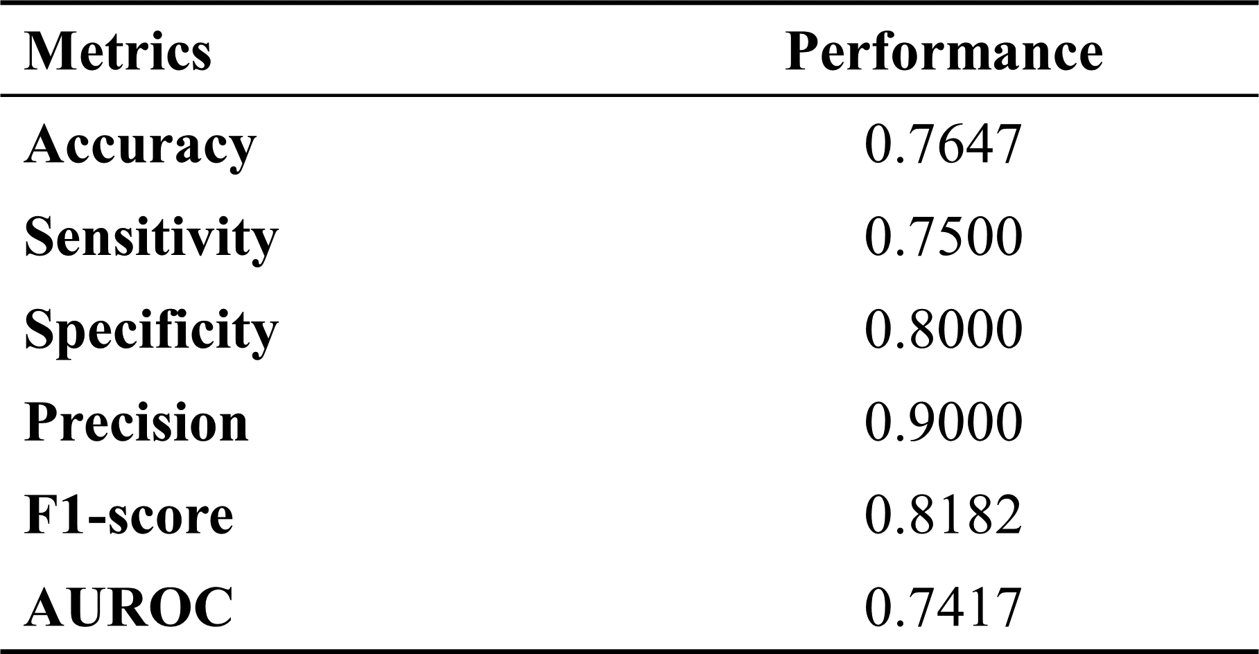
Results: A total of 165 patients were enrolled in the study. The mean age was 35.5 ± 12.47 years, and 63.7% were male. The cohort was predominantly HLA-B27 positive (93%), and 58.6% were classified as non-radiographic axSpA. At the time of imaging, 116 patients (70.7%) were clinically classified as having active disease. The proposed model was evaluated on a hold-out test set (randomly split 3:1). The deep learning model achieved an Area Under the Receiver Operating Characteristic Curve (AUROC) of 0.7417. The overall accuracy was 76.47%. In terms of specific diagnostic metrics, the model demonstrated a sensitivity of 75.00%, a specificity of 80.00%, a precision of 90.00%, and an F1-score of 0.8182, demonstrating a balanced detection capability.
Conclusions: We successfully developed an MRI-only deep learning model that effectively integrates features from the sacrum and spine to determine axSpA disease activity. By utilizing advanced cross-modal fusion techniques, the model demonstrated feasible performance to predict disease activity of patients with axSpA comparable to clinical judgment. This automated approach holds promise as an objective adjunctive tool in clinical practice, potentially mitigating inter-observer variability and facilitating standardized, quantitative assessment of disease activity in patients with axSpA.
Overview of the proposed multimodal deep learning framework for axial spondyloarthritis disease activity prediction using combined sacrum and spine MRI
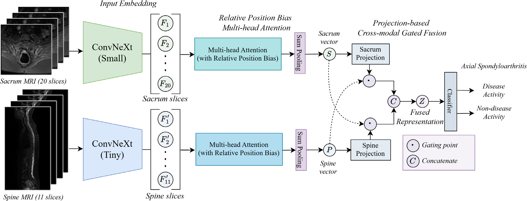
Table 1. Performance of the proposed MRI-only deep learning model for axial spondyloarthritis disease activity classification
REFERENCES: NIL.
Acknowledgments: NIL.
Disclosure of Interests: None declared.