fetching data ... 
Background: Giant cell arteritis (GCA) is a chronic granulomatous vasculitis driven by maladaptive immune responses and inappropriate tissue remodeling. While Tocilizumab (TCZ), an IL-6 receptor antagonist, effectively controls systemic inflammation and clinical manifestations of GCA, sequential biopsy studies reveal residual inflammation of the arterial wall [1]. Neutrophil extracellular traps (NETs), web-like chromatin structures, and senescent cells are specific inflammatory elements which accumulate in the arterial vascular wall of GCA patients, may persist for prolonged periods and serve as a source of IL-6 [2,3]. The impact of IL-6 receptor blockade on these elements and their contribution on persistent inflammation within the arterial wall remains unclear.
Objectives: To investigate the longitudinal impact of TCZ on the burden of NETs and senescent cells in sequential temporal artery biopsies (TABs) of GCA patients and explore the association with residual inflammation within the arterial vascular wall.
Methods: Sequential formalin-fixed paraffin-embedded (FFPE) temporal artery biopsy (TAB) sections from 11 GCA patients were analyzed pre- and post-TCZ treatment after a median TCZ treatment duration of 2.4 years (1.2 - 3.9). Immunofluorescence was employed to identify NETs (CitH3 + & MPO + DNA) and senescent cells (GL13 + , a biotinylated Sudan Black-B analogue specific for lipofuscin), simultaneously co-stained with IL-6, and infiltrating immune populations (CD3 + T-cells, CD20 + B-cells, CD68 + macrophages), followed by visualization via confocal microscopy. The ratios of total NETs and IL-6 + NETs volume relative to total tissue volume were quantified using the Imaris software. Neutrophil counts (MPO + ), GL13 + /IL-6 + senescent cells, CD3 + T-cells, CD20 + B-cells, CD68 + macrophages (immune infiltrates) were quantified using the Image J software. Clinical parameters (ESR, CRP, biopsy result, vascular PET, disease activity) were also correlated with the experimental findings. Δ median (%) was defined as follows: post-pre/pre %. Statistical analyses included Mann–Whitney and Wilcoxon tests, as well as Spearman correlations performed in GraphPad Prism and R package.
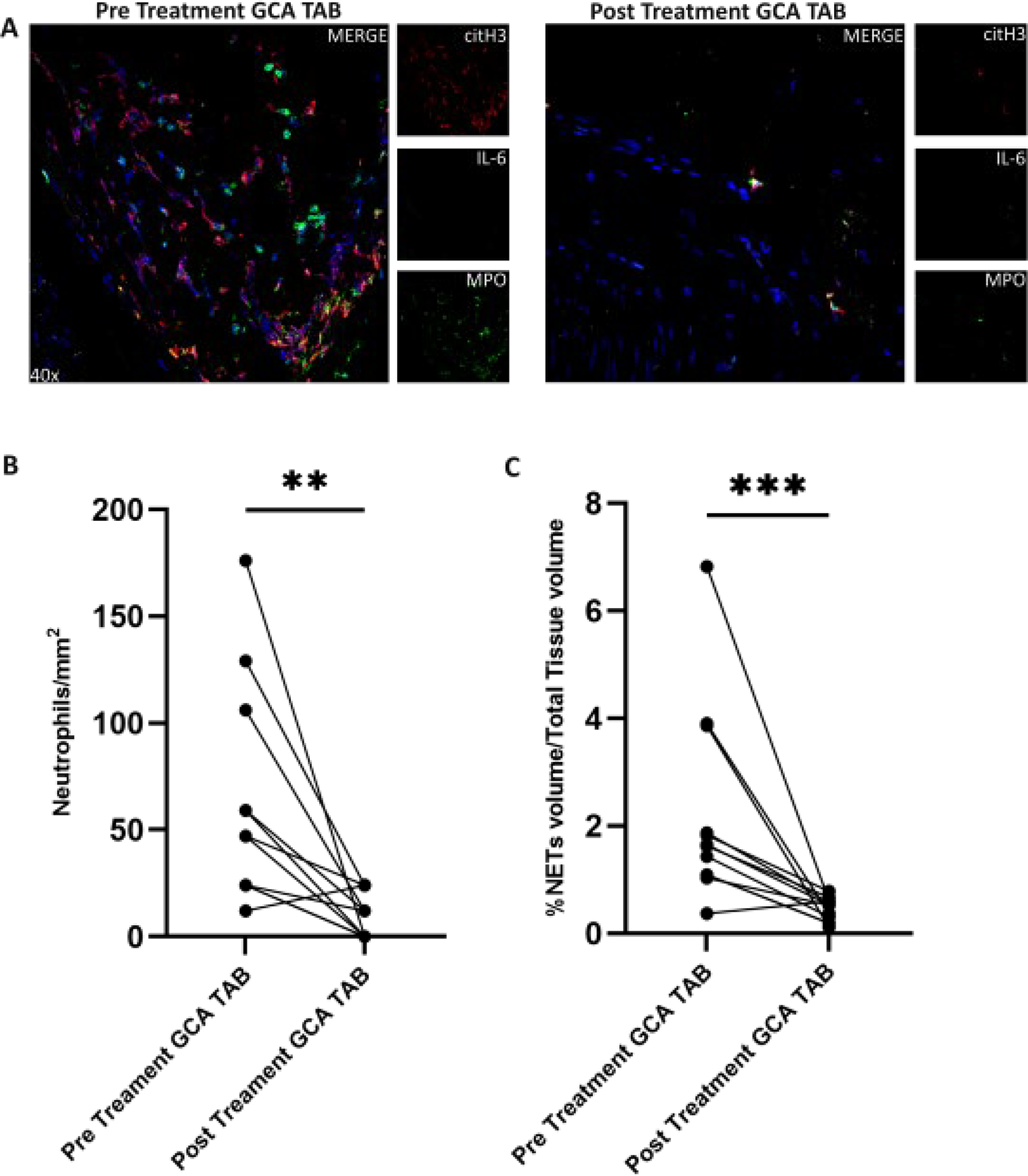
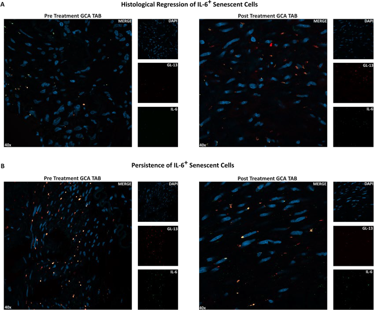
Results: After TCZ treatment, a statistically significant reduction in total NET volume fraction [pre vs post treatment median interquartile range (IQR) fraction (% ): 1.65 (1.34 - 2.86) vs 0.57 (0.45 - 0.64), p =0.001 ] ( Figure 1 ), IL-6 + senescent cells fraction [pre vs post treatment median (IQR) fraction (% ): 6.79 (2.59 - 7.51) vs 4.1 (1.75 - 5.5), p =0.03 ], neutrophil counts [pre vs post treatment median (IQR) cell counts ( cells/mm 2 ): 47 (24-82) vs 12 (0-18), p= 0.0029 ] and immune infiltrate counts [pre vs post treatment median (IQR) cell counts ( cells/mm 2 ): 870 (747-929) vs 35 (23-111), p = 0.02 ], was observed. Δ median (% ) was -67.9% , -30% , - 88.7% , and -93% for total NET volume fraction, IL-6 + senescent cells fraction, neutrophil counts and immune infiltrate counts, respectively. IL-6 + senescent cells exhibited a more modest Δ median reduction, with senescence-associated secretory phenotype (SASP) activity falling in 8 GCA patients but persisting at high levels in 3 ( Figure 2 ). There was not identified statistically significant correlation between Δ median (%) and either CRP or ESR. Likewise, no associations were identified between the experimental data and other clinical parameters (biopsy result, vascular PET, disease activity).
Conclusions: The long term treatment of GCA patients with anti-IL-6 receptor blockade leads to clinical remission and reduces the inflammatory bulk at the tissue level of the inflamed arterial vascular wall. However, NETs and senescent cells are only partially eliminated and may participate in disease relapses and future complications.
Tocilizumab (TCZ) reduces NETs and neutrophil counts in GCA Temporal Artery Biopsies (TABs). (A). Representative images from confocal microscopy showing NETs in TABs (n=22) of pre- and post-TCZ treated patients (n=11). Co-localization of extracellular DNA fibers with MPO (green) and citH3 (red) indicates the characteristic formation of NETs. (B) & (C). Change in percentage of NETs & neutrophil counts, respectively in TABs from GCA patients pre-post TCZ treatment. Each dot represents one patient. Lines connect paired samples.*** P < 0.001, ** P < 0.01, Wilcoxon signed-rank test. Scale bars= 20 μm
Variability in the Senescence-Associated Secretory Phenotype (SASP) following tocilizumab (TCZ) treatment. Representative Images from confocal microscopy of GCA patients Temporal Artery Biopsies (TABs) (n=22) showing the presence of senescent cells (GL-13, red) and IL-6 expression (green) pre- and post- TCZ treatment. (A) Histological regression of IL-6 + Senescent Cells: Upper panels illustrate a case where the absolute number of senescent cells remains stable post-treatment, but their pro-inflammatory IL-6 protein expression is significantly reduced in post treatment TABs (n=8). (B) Persistence of IL-6 + Senescent Cells: Lower panels illustrate a case where IL-6 protein expression does not change post treatment, indicative of a persistent inflammatory SASP phenotype of senescent cells in post-treatment TABs (n=3). Scale bars = 20 µm
REFERENCES: [1] Ricordi C, et al. RMD Open 2024;10:e005132.
[2] Palamidas DA, et al. Rheumatology 2022;61:1639-44.
[3] Veroutis D, et al. Ann Rheum Dis 2024;83:342-50.
Acknowledgments: NIL.
Disclosure of Interests: None declared.