fetching data ... 
Background: Takayasu arteritis (TAK) is a rare, chronic large-vessel vasculitis that primarily affects the aorta and its major branches. The disease predominantly occurs in Asian women under the age of 40, and its pathogenic mechanisms remain incompletely understood. Previous studies have indicated that multiple immune cell populations and their secreted cytokines contribute to vascular inflammation and structural damage in TAK, with CD4 + T cells, CD8 + T cells, and macrophages considered key drivers of disease pathology. In recent years, increasing attention has been paid to the role of CD8 + T cells in TAK, as they exhibit marked infiltration in both peripheral blood and affected aortic tissue and can mediate cytotoxic responses through the release of effector molecules such as Granzyme B. In addition, T cells can secrete chemokines that promote macrophage migration to inflamed vascular sites, while macrophages may further amplify inflammatory responses by producing chemokines such as CCL5 to recruit lymphocytes. However, the specific ligand–receptor pathways mediating interactions between CD8 + T cells and macrophages in TAK remain poorly defined.
Objectives: To characterize CD8 + T-cell heterogeneity in aortic tissue from patients with TAK using single-cell RNA sequencing (scRNA-seq), identify key CD8 + T-cell subsets with distinct molecular features, delineate their functional states, and explore their interaction networks with macrophages and other immune cell populations, thereby providing insights into the immune-pathogenesis of TAK and potential therapeutic targets.
Methods: Aortic tissue samples were obtained from three patients with TAK and three patients with atherosclerosis, all of whom met relevant clinical diagnostic and ethical criteria. Single-cell RNA sequencing was performed to profile the transcriptomes of aortic tissues. After standardized quality control and batch correction, major cell types were annotated, and TAK-derived CD8 + T cells were subjected to subclustering and functional characterization. Differential gene expression analysis, Gene Ontology (GO) and Kyoto Encyclopedia of Genes and Genomes (KEGG) enrichment analyses, and pseudotime trajectory inference were conducted to systematically assess functional heterogeneity and dynamic state transitions of CD8 + T cells. In addition, cross–cell-type pseudotime analyses integrating CD8 + T cells and macrophages were performed to infer potential temporal relationships. Cell–cell communication was further analyzed using the CellChat algorithm to identify ligand–receptor interaction networks between highly cytotoxic CD8 + T-cell subsets and other major cell types.
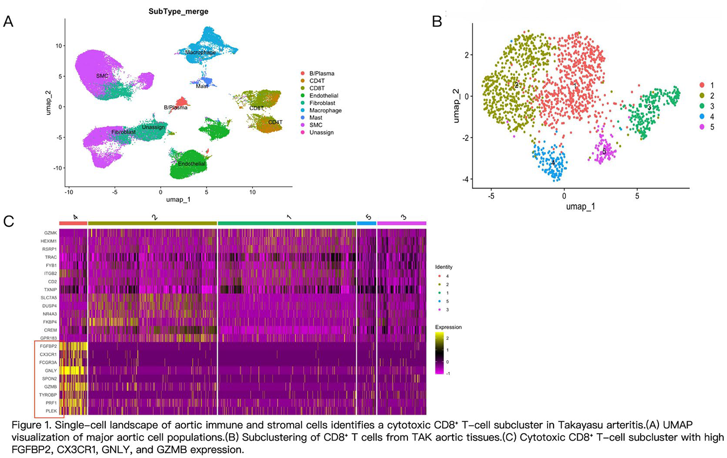
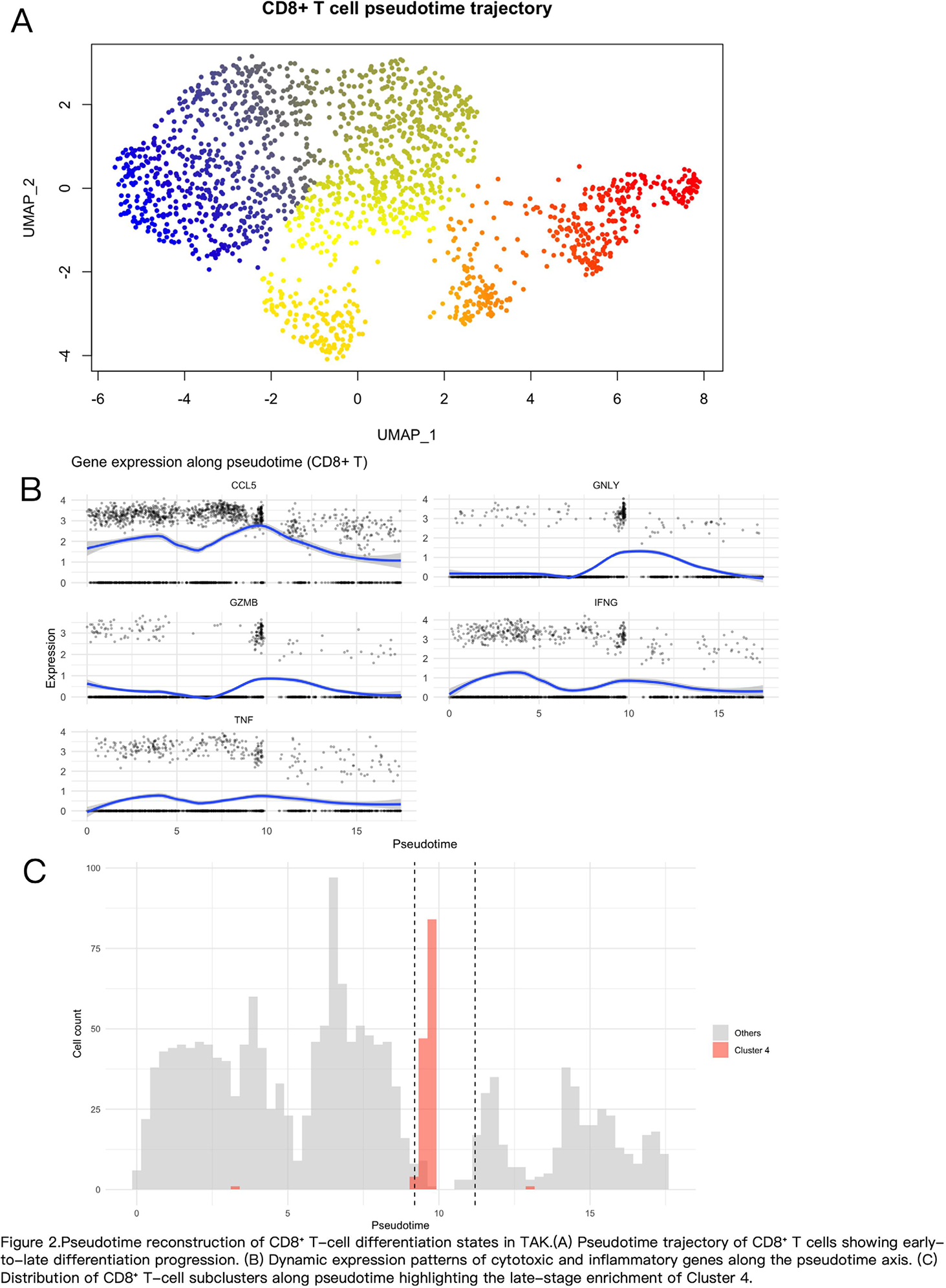
Results: Single-cell transcriptomic analysis of aortic tissue from patients with TAK revealed pronounced heterogeneity among CD8 + T cells and identified a key CD8 + T-cell subset with prominent cytotoxic features (cluster 4) (Figure 1). This subset was enriched for immune processes related to cell killing, inflammatory cytokine secretion, and chemotactic migration in GO and KEGG analyses. Pseudotime trajectory analysis placed this subset at a late activation stage, coinciding with peak expression of effector molecules including GZMB, GNLY, CCL5, and IFN-γ, suggesting a terminally differentiated, highly effector CD8 + T-cell state (Figure 2). Cell–cell interaction analysis further demonstrated that cluster 4 exhibited the strongest interaction intensity with macrophages, with chemokine-related signaling—particularly the CCL5–CCR1 axis—being most prominent. Integration of pseudotime analyses indicated that macrophage infiltration preceded CD8 + T-cell accumulation in affected aortic tissue, suggesting that macrophage-associated chemotactic signals may contribute to the enrichment of this highly cytotoxic CD8 + T-cell subset within inflammatory lesions.
Conclusions: Using single-cell RNA sequencing, this study delineates the transcriptional heterogeneity of CD8 + T cells in aortic tissue from patients with TAK and identifies a highly cytotoxic CD8 + T-cell subset characterized by late-stage activation. This subset is functionally associated with inflammatory responses, cytotoxic activity, and chemotaxis and exhibits prominent immune interactions with macrophages. Pseudotime analyses further suggest a dynamic temporal relationship between CD8 + T cells and macrophages during disease progression. These findings provide new insights into the coordinated roles of cytotoxic CD8 + T cells and innate immune cells in TAK and lay a foundation for further investigation into their functional significance and potential therapeutic targeting in large-vessel vasculitis.
REFERENCES: NIL.
Acknowledgments: NIL.
Disclosure of Interests: None declared.