fetching data ... 
Background: Takayasu arteritis (TAK) is a chronic granulomatous large-vessel vasculitis primarily involving the aorta and its main branches, and its pathogenesis remains unclear. Fibroblasts are important participants in vascular wall inflammation and remodeling.
Studies have shown that in the tumor microenvironment, cancer-associated fibroblasts (CAFs) can produce ligands such as FGF2 and FGF7 through autocrine or paracrine signaling. By activating FGFR signaling, these ligands help maintain the activated state of CAFs, thereby promoting tumor growth, angiogenesis, and immune evasion.
Objectives: This study aims to investigate the functions of various fibroblast subpopulations in Takayasu arteritis and to elucidate the mechanisms by which fibroblast-secreted FGF7 participates in vascular wall remodeling, thereby providing insights for the targeted therapy of TAK.
Methods: This study enrolled patients with Takayasu arteritis (TAK) from Beijing Anzhen Hospital, Capital Medical University, between January 2023 and October 2025. A total of 60 serum samples and 6 affected aortic tissue samples were collected for subsequent analysis. Additionally, serum samples from 20 healthy controls and 6 normal tissue samples from non-inflamed para-aortic sites were collected as controls. Serum FGF7 levels were measured using ELISA in the 60 TAK patients and 20 healthy controls. Single-cell RNA sequencing (scRNA-seq) and proteomic analyses were performed on aortic tissue samples from three TAK patients and three controls.
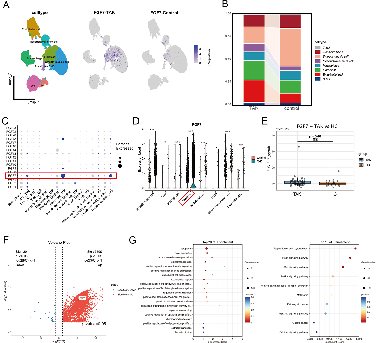
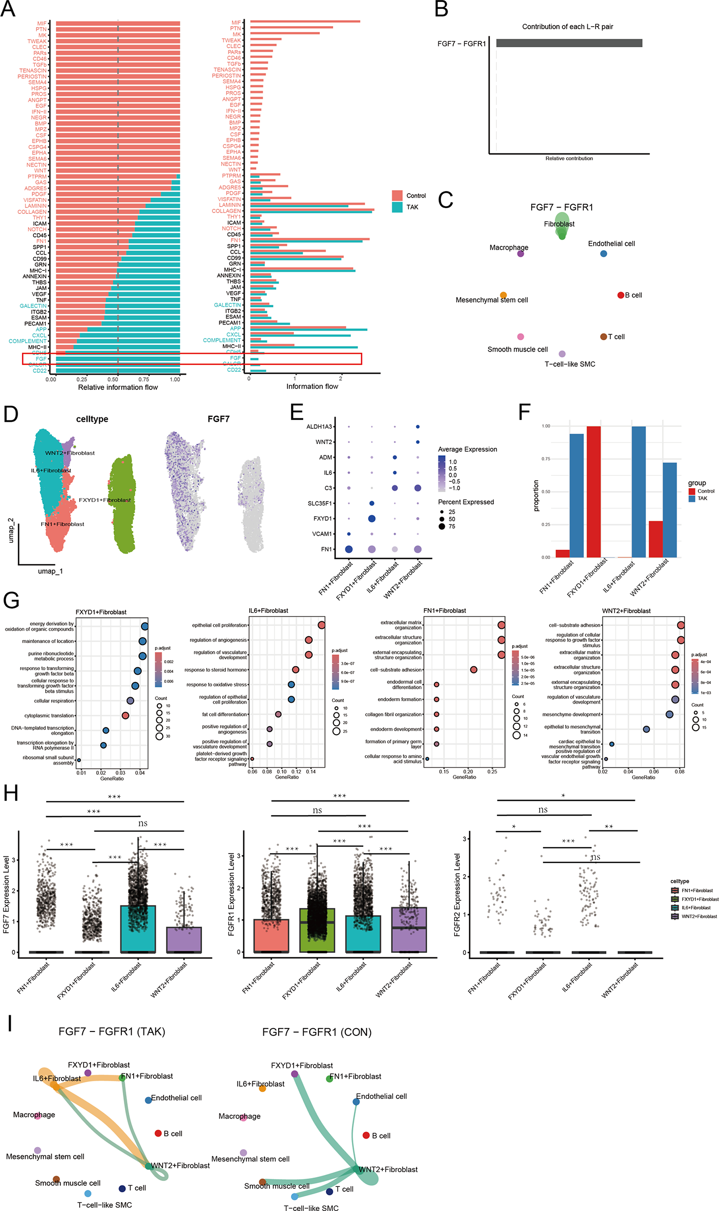
Results: ScRNA-seq and Proteomics Reveal Significant Upregulation of FGF7 in Patients with TAK ScRNA-seq of aortic specimens from three TAK patients and three healthy controls resolved eight transcriptionally distinct subsets: smooth muscle cells (SMCs), T cells, macrophages, fibroblasts, endothelial cells, B cells, mesenchymal stem cells, and a rare T-cell-like SMCs population ( Figure 1A ). The number of fibroblasts in the aortic tissue of TAK patients was significantly increased compared to the control group (5,794 vs. 4,129) ( Figure 1B ). Expression profiling of FGF family members showed that not only were more fibroblasts positive for FGF7 in TAK, but the FGF7 transcript level per fibroblast was also markedly elevated compared with controls (p < 0.001) ( Figure 1C-D ). No significant difference in serum FGF7 levels was observed between the TAK group and the control group (p=0.46), which may be attributed to the autocrine or paracrine mode of action of FGF7 ( Figure 1E ). Proteomic analysis of aortic tissues from three patients with TAK and three controls demonstrated a significant upregulation of FGF7 in the TAK group [P < 0.05, log2(FC) > 1] ( Figure 1F ). GO (Gene Ontology) and KEGG (Kyoto Encyclopedia of Genes and Genomes) enrichment analyses were performed, showing the top 20 GO terms and top 10 KEGG pathways that encompass FGF7 ( Figure 1G ). Upregulation of FGF7-FGFR1 Signaling Between Fibroblasts in TAK CellChat analysis of the transcriptomic data revealed that FGF signaling was predominantly active in the TAK group ( Figure 2A ). The FGF7-FGFR1 ligand-receptor pair was identified as the primary contributor to this pathway, primarily mediating communication between fibroblasts ( Figure 2B-C ). This study found that although FGFR2 is the canonical receptor for FGF7, its expression is minimal in the aortic tissues of both TAK and control groups. In contrast, the expression of the non-canonical receptor FGFR1 is significantly higher than that of FGFR2. Moreover, both overall and fibroblast-specific expression of FGFR1 are markedly upregulated in the TAK group.
IL6+ Fibroblasts Exhibit Marked Upregulation of FGF7 and FGFR1 After extracting the fibroblasts, we performed dimensionality reduction and clustering again. Based on the gene expression patterns of the four clusters, they were annotated as FXYD1+ fibroblasts, IL6+ fibroblasts, FN1+ fibroblasts, and WNT2+ fibroblasts ( Figure 2D ). Notably, over 99% of IL6+ fibroblasts were distributed within the TAK group ( Figure 2F ). FGF7 expression in IL6 + fibroblasts is significantly higher than in other clusters. Meanwhile, IL6 + and FN1 + fibroblasts—predominantly distributed in the TAK group—exhibit markedly elevated FGFR1 expression compared to the other two clusters ( Figure 2H ). This suggests that in TAK, IL6 + fibroblasts may produce FGF7 through autocrine/paracrine signaling, subsequently activating other fibroblast subpopulations via FGFR1 binding. CellChat analysis further revealed that, compared to controls, the FGF7-FGFR1 ligand–receptor pair primarily mediates communication between IL6 -+ - fibroblasts and IL6 -+ -, FN1 -+ -, and WNT2 -+ - fibroblasts within the TAK group ( Figure 2I ). GO enrichment analysis performed separately for each subpopulation revealed distinct functional profiles: IL6 + fibroblasts were primarily enriched in angiogenesis-related processes such as regulation of angiogenesis and vasculature development; FN1 + fibroblasts showed significant enrichment in extracellular matrix remodeling processes including extracellular matrix organization and cell-substrate adhesion; while WNT2 + fibroblasts were mainly enriched in processes such as cell-substrate adhesion and extracellular matrix organization ( Figure 2G ).
Conclusions: In this study, by integrating transcriptomic and proteomic analyses of aortic tissues from both the TAK group and the control group, we have for the first time proposed that FGF7 is upregulated in the aortic tissues of TAK patients. Furthermore, IL6+ fibroblasts are capable of producing FGF7 in an autocrine or paracrine manner and, through binding to its receptor FGFR1, can activate other fibroblast subpopulations. On one hand, this process may positively regulate angiogenesis within the arterial wall; on the other hand, it may promote extracellular matrix remodeling, ultimately contributing to vascular wall remodeling in Takayasu arteritis.
REFERENCES: NIL.
Acknowledgments: NIL.
Disclosure of Interests: None declared.