fetching data ... 
Background: Familial Mediterranean fever (FMF) is a pyrin-inflammasome–driven autoinflammatory disease characterized by recurrent inflammatory flares. While disease models largely emphasize myeloid cytokine amplification, the contribution of cytotoxic lymphocytes remains insufficiently defined. In systemic autoinflammatory disorders, natural killer (NK) cells are increasingly recognized as inflammation-sensitive effectors at the interface of innate sensing and immune regulation, yet their role is poorly documented across AIDs. Recent evidence in related autoinflammatory settings supports that NK cells can acquire states of quantitative loss, activation–exhaustion, apoptosis susceptibility and impaired cytokine output, suggesting that NK dysfunction may represent a broader and clinically meaningful axis of immune dysregulation.
Objectives: To characterize NK-cell deficiency and stress programs during FMF attacks using integrated multi-omics profiling, and to identify inflammatory pathways associated with NK-cell dysfunction.
Methods: We analyzed 38 genetically confirmed FMF patients sampled during flares and 68 healthy controls from the ImmunAID European cohort. Multi-layer profiling comprised bulk PBMC RNA-seq, single-cell RNA-seq, miRNA-seq of sorted immune subsets, high-parameter NK flow cytometry, soluble inflammatory mediators, SomaScan plasma proteomics, and targeted lipid mediator quantification. We integrated flare-associated changes across mRNA, cellular and surface protein phenotypes, and systemic fluids (plasma/urine), projected signals onto PBMC subtypes, and validated key modules through cross-omics concordance and clinico-biological correlation analyses.
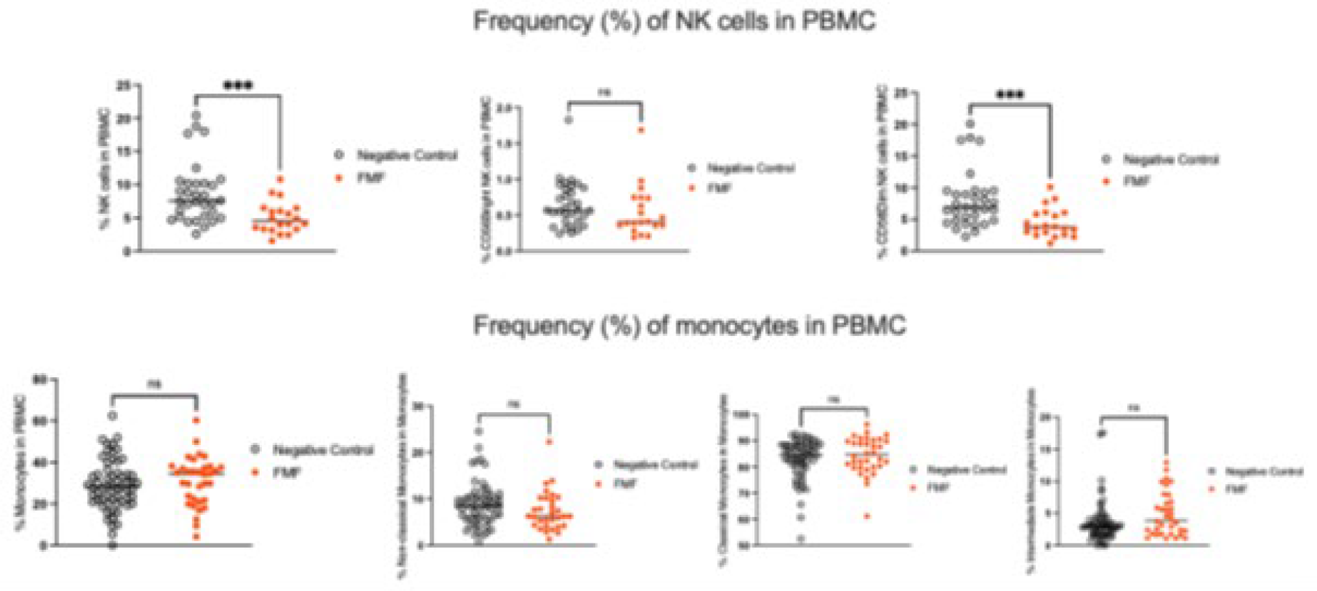
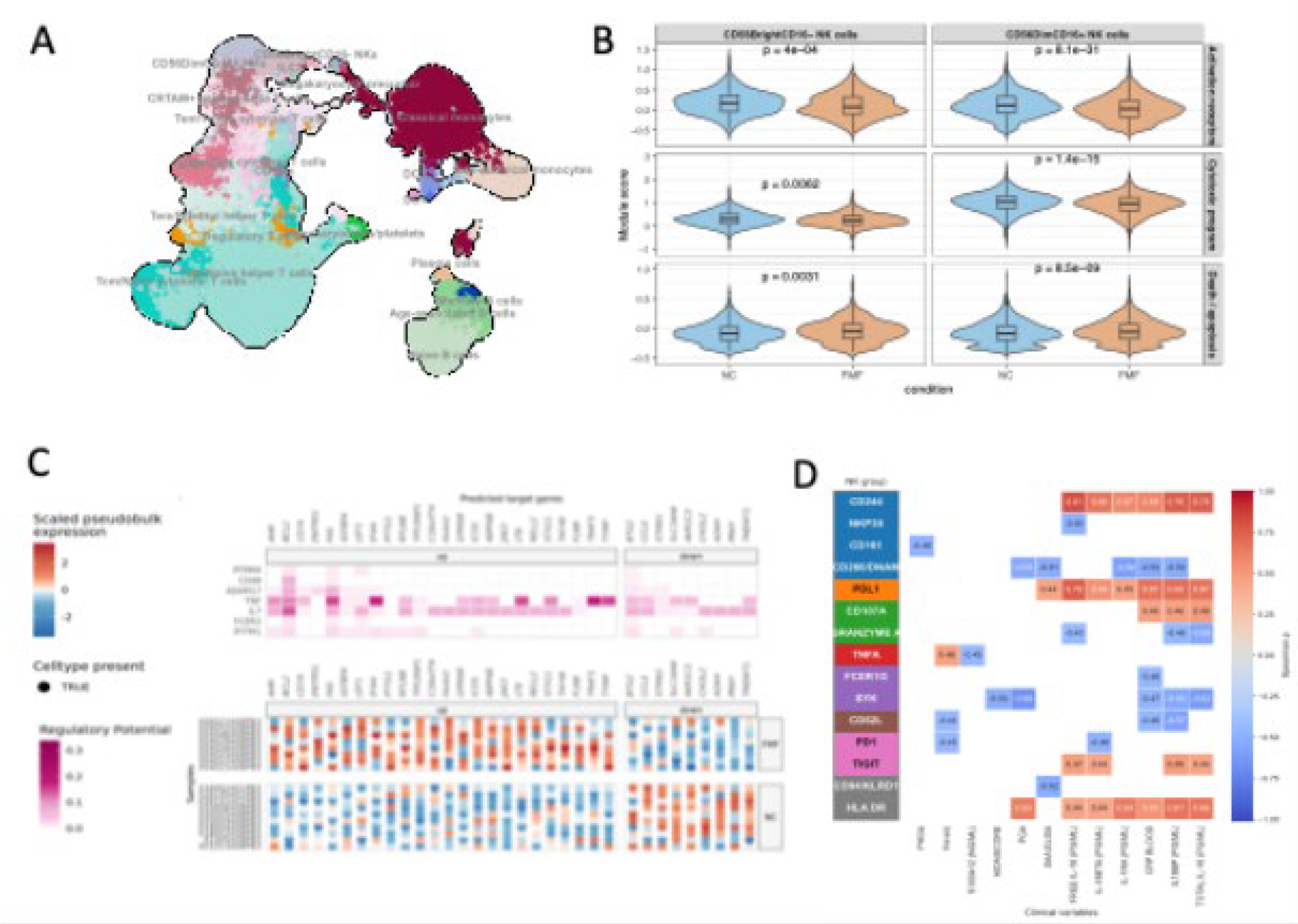
Results: Bulk PBMC RNA-seq identified 106 differentially expressed genes (44 upregulated, 62 downregulated) in FMF versus controls, highlighting innate inflammatory activation (including IL1B, CXCL8) together with marked suppression of NK/T cytotoxic programs (GZMB, PRF1, KIR2DL1/3, CCL3) and reduced NK identity regulators (TBX21, ZBTB16/PLZF). Pathway and regulon analyses indicated TNF signaling activation with repression of IFN–JAK–STAT and TGF-β programs. At the systemic level, soluble mediator profiling and SomaScan plasma proteomics revealed a coordinated cytokine–complement inflammatory milieu during attacks, with convergent enrichment of pathways linked to TNF/IL-1–driven inflammation, complement activation, and tissue stress, consistent with transcriptome-derived upstream regulators. Targeted lipid mediator profiling further demonstrated flare-associated remodeling of bioactive lipid networks, supporting an activated inflammatory state. A prespecified 10-gene NK/IL-1/ECM panel, dominated by cytotoxic and trafficking genes together with IL-1/ECM-linked components, discriminated FMF flares from controls with high accuracy (Random Forest 88%, XGBoost 85%) and outperformed alternative gene signatures. Flow cytometry confirmed a significant reduction of total NK cells during flares (4.5% vs 7.58%, P=0.0004), driven by selective depletion of CD16 + CD56Dim cytotoxic NK cells (3.7% vs 6.9%, P=0.003) ( Figure 1 ), whereas CD16 − CD56Bright NK cells were preserved. Single-cell RNA-seq supported predominant perturbation of the NK compartment, with CD16 + CD56Dim NK cells exhibiting stress/apoptosis skewing and reduced cytotoxic mediators ( Figure 2 ), consistent with a TNF-linked attrition program coupled to activated myeloid inflammatory circuits. Across omics layers, NK deficiency/stress markers coherently tracked with systemic inflammatory signatures and were associated with FMF clinical manifestations and flare severity, supporting NK-cell dysfunction as a potential stratification axis and biomarker framework in FMF.
Conclusions: FMF flares are characterized by selective depletion and stress-associated rewiring of cytotoxic CD16 + CD56Dim NK cells, with relative preservation of CD16 - CD56Bright NK cells, within a TNF/IL-1–skewed cytokine–complement–lipid inflammatory milieu. A compact NK/IL-1/ECM signature robustly discriminates FMF flares from controls and suggests NK dysfunction as a clinically relevant stratification axis with potential utility for biomarker-guided monitoring and pathway-informed therapeutic evaluation.
Flow-cytometric enumeration of NK- and monocyte subsets in FMF flares and controls.
PBMC from FMF flare patients and age/sex-matched non-inflammatory controls. PBMCs were isolated within 2 h by Ficoll, cryopreserved, thawed, stained with a 14-colour antibody panel (CD3, CD19, CD14, CD16, CD56, HLA-DR, CD11c, CD45, viability dye), and acquired on flow cytometry. Doublets and dead cells were excluded. NK cells were defined as CD3 − CD19 − CD14 − CD56 + and subdivided into CD56Bright (Immunoregulatory and CD56Dim(cytotoxic) subsets. Monocytes were defined as CD14 + HLA-DR + and subdivided into classical (CD14 + CD16 − ), intermediate (CD14 + CD16 + ), and non-classical (CD14 + CD16 + ) subsets. Frequencies are displayed as percentages of live PBMCs (total NK cells, total monocytes) or of the parent population (monocyte subsets).
Multi-omics evidence for selective cytotoxic NK-cell loss during FMF flares.
(A) Single-cell PBMC atlas (UMAP) identifying major immune subsets, with focus on the NK-cell compartment.
(B) Preferential depletion of cytotoxic NK cells during flares: violin plots show a marked reduction of CD56^dim^CD16
+ NK cells in FMF flares versus controls, while CD56^bright^CD16
− NK cells are relatively preserved.
(C) NK dysfunction program combining effector loss and regulatory rewiring: heatmaps display altered expression of key NK effector/identity genes and inferred regulatory activity across PBMC subsets.
(D) NK-associated signatures correlate with inflammatory burden: correlation heatmap links NK stress/deficiency markers to clinical and biological indicators of FMF activity and severity (red positive, blue negative associations).
REFERENCES: NIL.
Acknowledgments: NIL.
Disclosure of Interests: None declared.