fetching data ... 
Background: Psoriatic arthritis (PsA) is an immune-mediated chronic inflammatory arthropathy in which Th17 cells and Th17 cytokines (IL-17, IL-21 and IL-22) play a prominent role. Semaphorin4A (Sema4A), is a protein that belongs to the Semaphorin protein family. Sema4A is a key molecule in the regulation of T cell homeostasis, activation, and Th17 differentiation and is implicated in the pathogenesis of several rheumatic diseases, including rheumatoid arthritis and systemic sclerosis.
Objectives: The aim of this work was to determine the expression of Sema4A in patients with PsA and to analyze the functional outcomes of Sema4A activation both in vitro and in vivo .
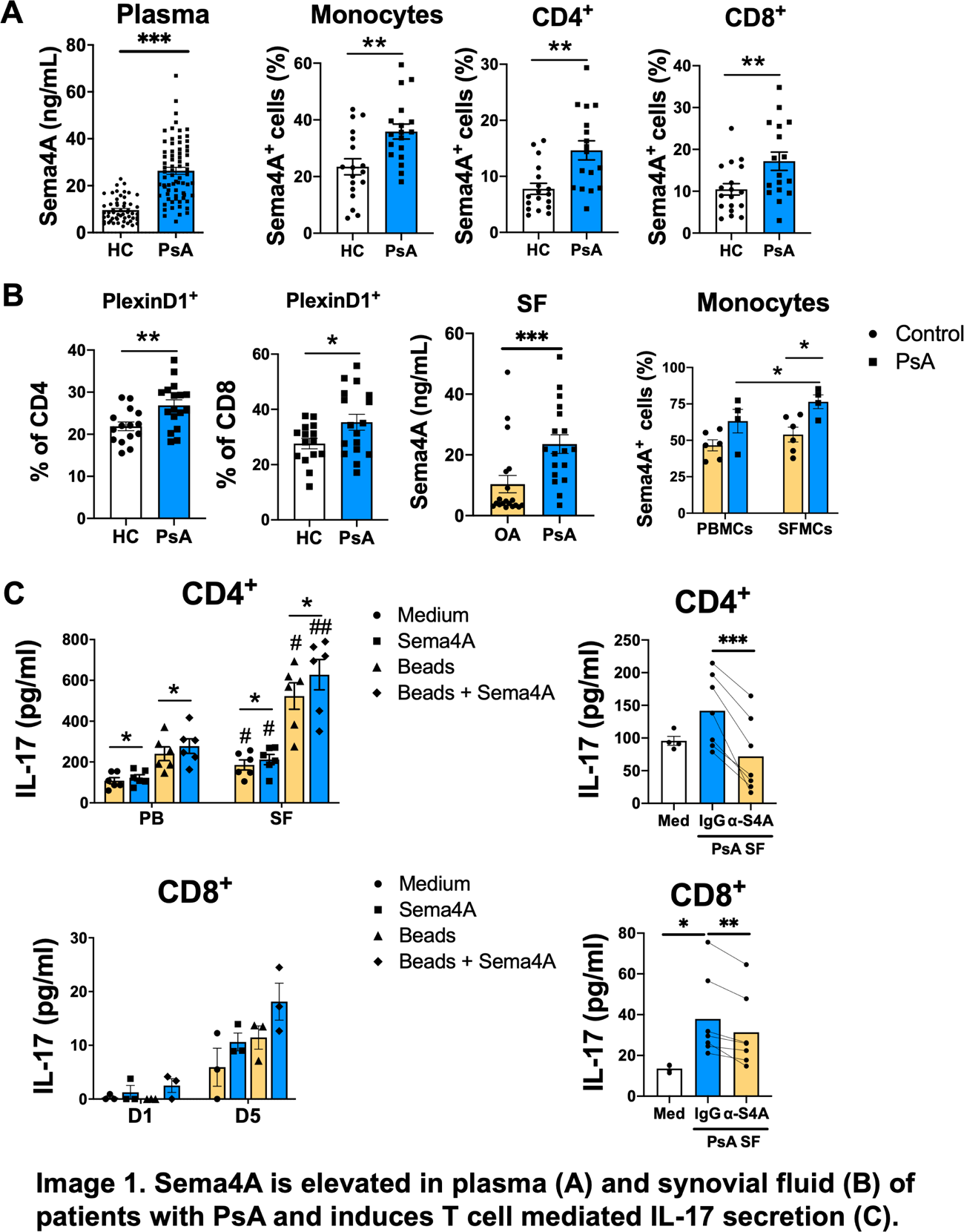
Methods: Plasma and synovial fluid (SF) levels of Sema4A were measured by ELISA. Expression of Sema4A and its receptors PlexinB2, PlexinD1 and Neuropilin-1 was determined by flow cytometry in peripheral blood mononuclear cells (PBMC) and SF mononuclear cells (SFMC). CD4 + and CD8 + T cells were stimulated with anti-CD3/anti-CD28 beads (ratio 1 bead: 5 cells) alone or in combination with recombinant human Sema4A (200 ng/mL), or with PsA SF (25 % v/v) in the presence of an anti-human Sema4A antibody or its respective IgG isotype control (both 1 µg/mL). In vivo, we used the Collagen Induced Arthritis (CIA) model, in which mice were treated with an anti-Sema4A neutralizing antibody or its respective IgG isotype control (both 100 µg/mouse). mRNA and protein expression of inflammatory mediators was analyzed by RT-qPCR, ELISA and flow cytometry
Results: Plasma and SF levels of Sema4A were significantly elevated in patients with PsA compared to healthy controls (HC) and patients with osteoarthritis (OA), respectively. Moreover, the frequency of PB and SF monocytes, CD4 + and CD8 + T cells expressing Sema4A was significantly higher in PsA than in HC and OA, as well as the percentage of CD4 + and CD8 + T cells expressing PlexinD1. Functionally, Sema4A induced mRNA and protein expression of Th17 cytokines by CD4 + and CD8 + T cells from both PB and SF. Importantly, the SF of PsA patients induced the secretion of IL-17 by non-stimulated and CD3/CD28-stimulated T cells from HC, but the addition of an anti-Sema4A antibody drastically abolished this secretion, demonstrating that Sema4A present in the SF of PsA patients induces the production of IL-17.
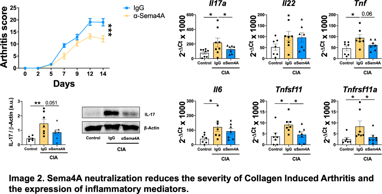
Finally, the administration of an anti-Sema4A neutralizing antibody reduced the severity of CIA, which was associated with a reduced mRNA expression of Il17a , Tnf , Tnfsf11 (encoding RANKL) and Tnfrsf11a (encoding RANK) genes and a reduced IL-17 protein expression in the joints of the anti-Sema4A treated mice.
Conclusions: This study demonstrates that Sema4A signaling is deregulated in patients with PsA and that Sema4A plays an important role in IL-17-mediated inflammation. Therefore, Sema4A could be a promising therapeutic target for the treatment of PsA and other IL-17-mediated diseases.
REFERENCES: NIL.
Acknowledgments: NIL.
Disclosure of Interests: None declared.