fetching data ... 
Background: 3H-1,2-Dithiole-3-Thione (D3T), a cruciferous vegetable–derived antioxidant and activator of the Nrf2 transcription pathway, has demonstrated immunomodulatory effects in murine models of experimental autoimmune encephalomyelitis (EAE) and psoriasis [1,2]. In EAE, D3T inhibited pathogenic Th1 and Th17 cell differentiation [1], and in psoriasis model it suppressed pathogenic Th17 cell differentiation [2]. Because Th1 and Th17 cells are among the central drivers of autoantibody production and tissue injury in systemic lupus erythematosus (SLE) [3,4], the therapeutic potential of D3T in lupus warrants investigation
Objectives: To evaluate the therapeutic effect of D3T on lupus nephritis and to investigate its immunomodulatory actions in the imiquimod (IMQ)-induced lupus-prone mouse model.
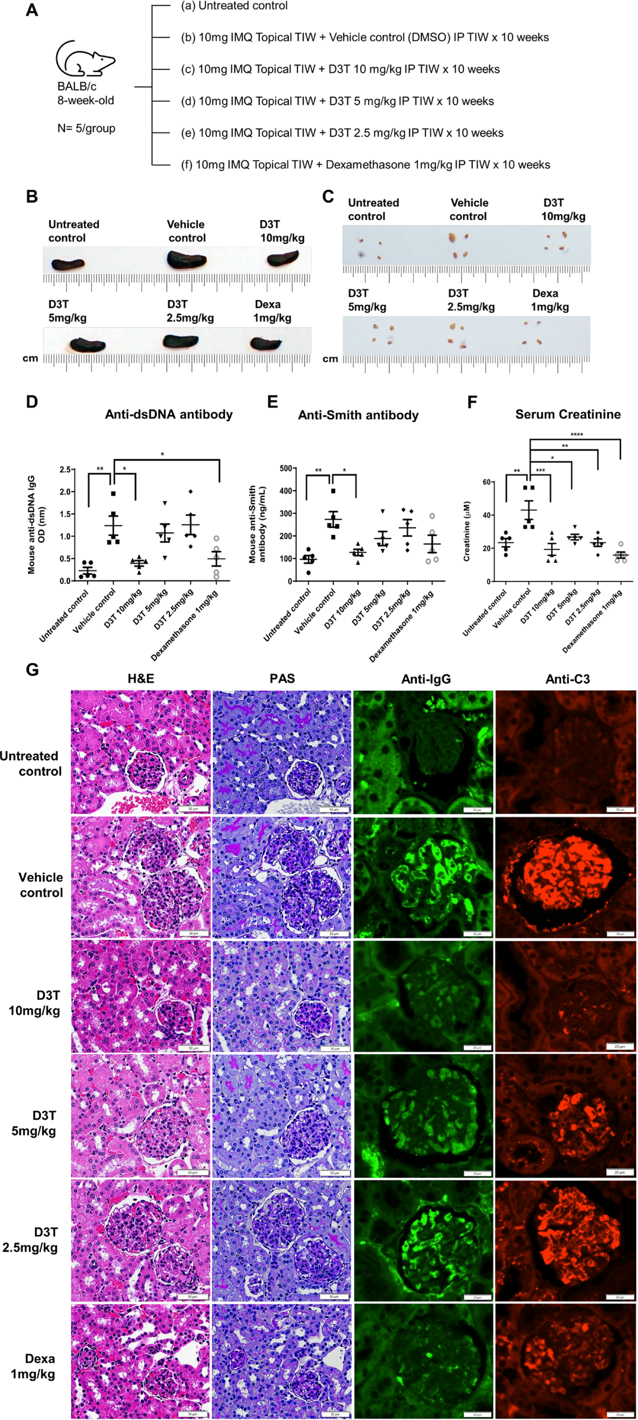
Methods: Eight-week-old BALB/c mice (n = 5 per group) were treated topically with IMQ cream (10 mg), a Toll-like receptor 7 agonist, on the right ear three times per week for 10 weeks to induce lupus-like manifestations (Figure 1A). D3T at doses of 2.5, 5, and 10 mg/kg was administered intraperitoneally three times per week for 10 weeks. Control groups included untreated mice, vehicle-treated mice (DMSO), and a positive control group receiving dexamethasone (1 mg/kg, intraperitoneally) on the same schedule. At week 10, mice were sacrificed for analysis. Serum autoantibodies and cytokines were quantified by enzyme-linked immunosorbent assay (ELISA). Renal histopathology was evaluated using hematoxylin and eosin (H&E), Periodic Acid-Schiff (PAS), and immunofluorescence staining. Th cell subsets in spleen and cervical lymph nodes were analyzed by flow cytometry
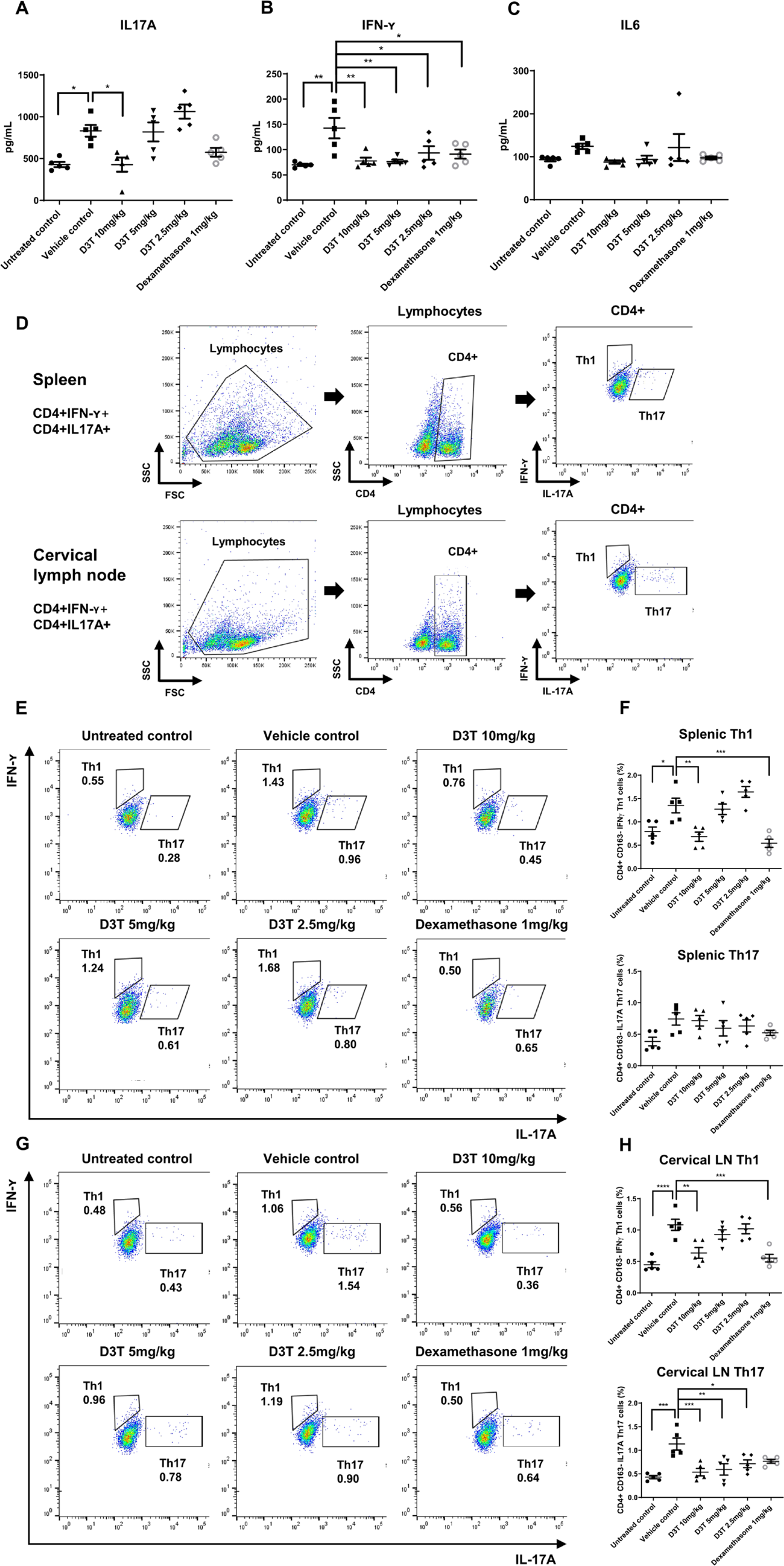
Results: D3T treatment reduced splenomegaly and cervical lymphadenopathy in IMQ-induced lupus-prone mice (Figure 1B and 1C). Serum anti-dsDNA and anti-Smith antibody levels decreased in a dose-dependent manner (Figure 1D and 1E), and serum creatinine remained stable in D3T- and dexamethasone-treated groups (Figure 1F). Histological and immunofluorescent analyses demonstrated that D3T prevented the development of proliferative glomerulonephritis and glomerular immune complex deposition (Figure 1G). D3T significantly reduced serum IL-17A and IFN-γ levels, while IL-6 remained unaffected (Figure 2A, 2B and 2C). Flow cytometric analysis (Figure 2D) revealed that D3T reduced the proportions of Th1 cells in spleen and cervical lymph nodes (Figure 2E and 2F), and Th17 cells in cervical lymph nodes (Figure 2G and 2H).
Conclusions: This study provides the first evidence that D3T ameliorates lupus-like disease in mice. D3T decreased lupus-specific autoantibody production, alleviated renal inflammation, and inhibited differentiation of Th1 and Th17 cells. These findings suggest that D3T is a promising therapeutic candidate for SLE and lupus nephritis. Further mechanistic studies are warranted.
D3T ameliorates lupus-like manifestations in IMQ-induced lupus-prone mice. (A) Experimental design of animal study. Representative photographs of (B) spleens and (C) cervical lymph nodes at week 10. Serum levels of (D) anti-dsDNA antibodies, (E) anti-Smith antibodies, and (F) creatinine are shown. Error bars indicate the standard error of the mean (SEM). P values were calculated using one-way ANOVA (*p < 0.05, **p < 0.01, ***p < 0.001, ****p < 0.0001). (G) Representative kidney sections stained with H&E and PAS (scale bars = 50 µm), and immunofluorescence staining for IgG and C3 deposition (scale bars = 20 µm).
D3T inhibits production of IL-17A and IFN-γ and reduces Th1 and Th17 cell frequencies. Serum levels of (A) IL-17A, (B) IFN-γ, and (C) IL-6 at week 10 are shown. (D) Gating strategy for flow cytometric analysis of Th cell subsets. Th17 cells were defined as CD4 + IL-17A + IFN-γ − cells, and Th1 cells as CD4 + IL-17A − IFN-γ + cells. (E) Representative plots and (F) quantitative analysis of Th1 and Th17 cells in the spleen. (G) Representative plots and (H) quantitative analysis of Th1 and Th17 cells in cervical lymph nodes. Error bars indicate the SEM. P values were calculated using one-way ANOVA (*p < 0.05, **p < 0.01, ***p < 0.001, ****p < 0.0001).
REFERENCES: [1] Kuo PC, Brown DA, Scofield BA, et al. 3H-1,2-dithiole-3-thione as a novel therapeutic agent for the treatment of experimental autoimmune encephalomyelitis. Brain Behav Immun. 2016;57:173-86.
[2] Shih MC, Li CL, Liao EC, et al. Inhibition of NLRP3 Inflammasome Activation by 3H-1,2-Dithiole-3-Thione: A Potential Therapeutic Approach for Psoriasis Treatment. Int J Mol Sci. 2023;24(17):13528.
[3] Tang YY, Wang DC, Chen YY, et al. Th1-related transcription factors and cytokines in systemic lupus erythematosus. Front Immunol. 2023 Dec 18;14:1305590.
[4] Hoi A, Igel T, Mok CC, et al. Systemic lupus erythematosus. Lancet. 2024;403(10441):2326-38.
Acknowledgments: NIL.
Disclosure of Interests: None declared.