fetching data ... 
Background: The previously established Knee Inflammation MRI Score (KIMRISS) interface incorporates slice-by-slice dichotomous scoring of bone marrow lesions (BML) on sagittal knee MRI in 28 regions defined by grid templates manually placed on the patella, femur, and tibia. KIMRISS BML scoring demonstrates high inter-rater reliability and sensitivity to small longitudinal changes in lesion volume [1]. While the established platform is relatively user-friendly and provides consistency for scoring input, recent advances in artificial intelligence algorithms could greatly decrease the amount of human time and effort required to assess knee BML by automatically identifying scoring regions and evaluating images for the presence of lesions.
Objectives: We aimed to develop a BML detection algorithm that uses the 28-region KIMRISS grid framework to produce an automated semi-quantitative knee BML score (“iKIMRISS”) comparable to scores manually generated by calibrated human experts.
Methods: First, automatic anatomy-conforming grid placement methods were developed via geometric analysis of bone masks (annotated MR images) applied to the femoral condyles, tibia, and patella. Femur grids were generated by using least squares to fit an ellipse to the femoral mask and project radial lines from the center point of each condyle to the outer edges at equal angles. Bounding boxes were fitted to the tibial and patellar edges. The width of the tibial bounding box was used to divide the region into a maximum of 5 equal sections from anterior to posterior, which were each further subdivided into two 1cm superior and inferior regions. The height of the patellar bounding box was used to divide the region of interest into a maximum of 4 equal sections. Second, a 3D segmentation model based on an in-context learning model known as Temporal, was trained on 150 sagittal knee MRIs annotated for bone marrow lesions. The training dataset included images from the OKINADA adalimumab trial, the publicly available Osteoarthritis Initiative dataset, and anonymized images obtained from our local hospital PACS system. Finally, the automated grid placement and BML segmentation methods were applied to n=40 new cases with baseline and follow-up scans (n=20 from the OKINADA trial, baseline and 16 week follow-up; n=20 from the publicly available Osteoarthritis Initiative Dataset). The data from the two algorithms were combined and processed to produce a dichotomous (0/1) BML score for each grid region, and the grid scores were added together to produce a total semi-quantitative iKIMRISS score (Figure 1).
To evaluate scoring reliability, intra-class correlation coefficients (ICCs) were calculated between the fully automated iKIMRISS scores and manual scores generated by two expert readers in previous exercises.
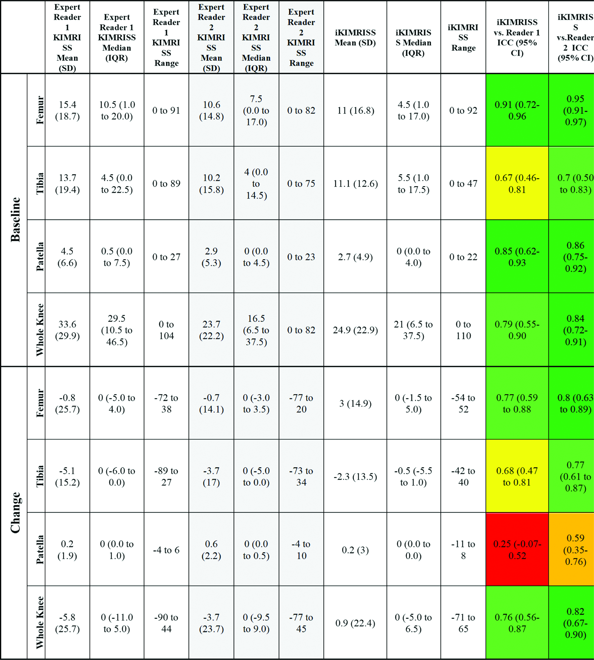
Results: Intra-class correlation coefficients for human vs. automated scores are shown in Table 1. Reliability of iKIMRISS BML status scores was very good for the femur and patella (ICC >0.90), moderate for the tibia (ICCs 0.65 and 0.70), and good when considering the whole knee (ICCs 0.78 and 0.84). Reliability for iKIMRISS BML change scores was good in the femur, the whole knee, and one reader’s tibial scores (ICCs 0.76-0.82). Reliability with the other reader’s tibial change score was moderate (ICC 0.68), and lowest reliability was seen for both readers’ patella change scores (ICCs 0.25 and 0.59).
Conclusions: The novel iKIMRISS grid placement and BML detection algorithm demonstrates good reliability with expert human reader KIMRISS scores. Lower ICCs for patella change scores may be due, in part, to a relatively low frequency of change observed in the patella in this dataset.
An illustrative sample of fluid-sensitive sagittal knee MRI slices taken from a single case with (right column) and without (left column) iKIMRISS annotation. Bone-contouring KIMRISS grid templates for the femur, tibia, and patella are shown in orange, cyan, and pink, respectively. Bone marrow lesion segmentations in the femur and patella are shown in red and green, respectively.
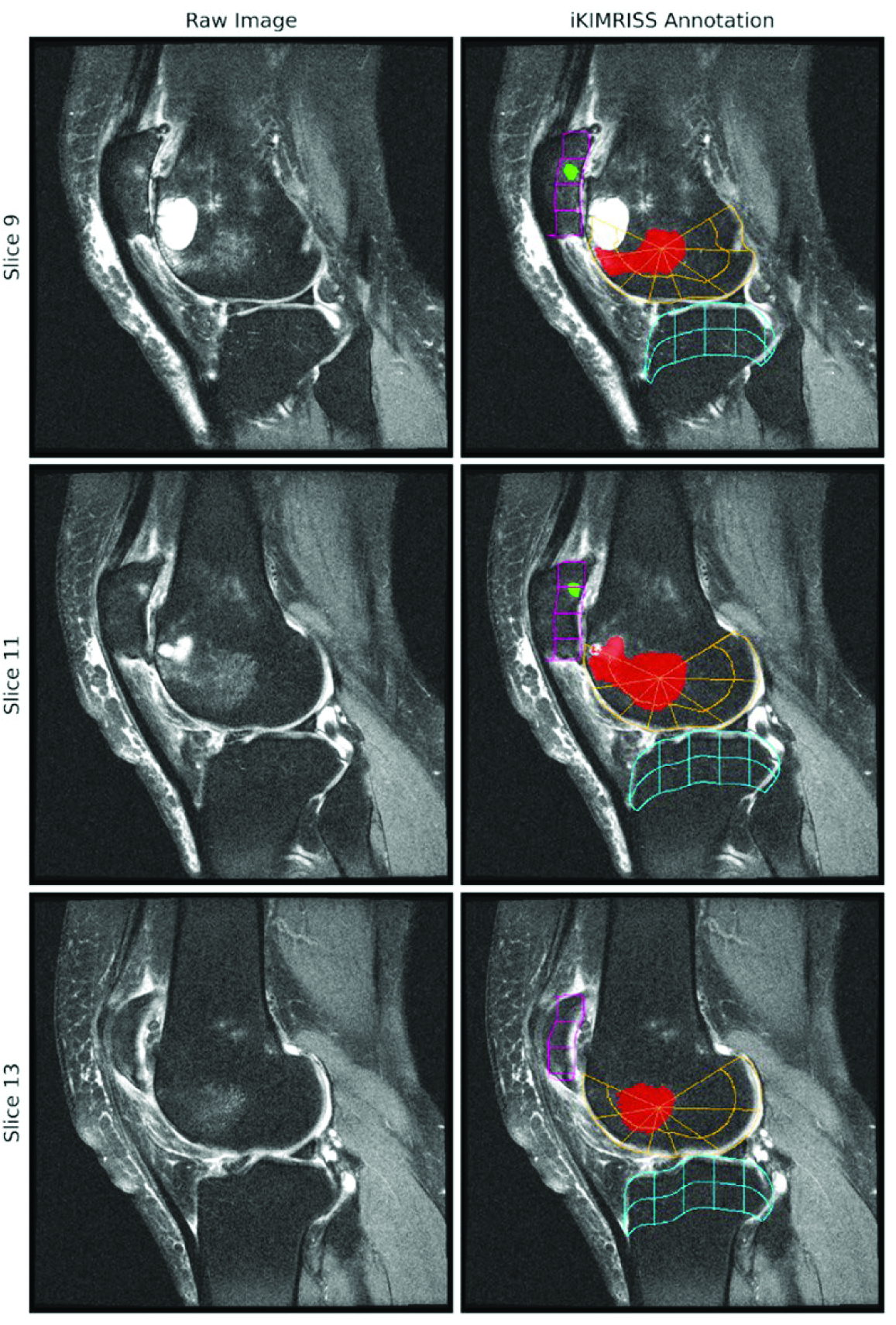
Table 1. Descriptive data and reliability for bone marrow lesions in n=40 osteoarthritis patients from the Osteoarthritis Initiative Dataset and OKINADA adalimumab trial, evaluated via manual KIMRISS scoring by two human expert readers and the automated iKIMRISS BML detection algorithm.
REFERENCES: [1] Maksymowych WP, Jaremko JL, Pedersen SJ, et al. Comparative validation of the knee inflammation MRI scoring system and the MRI osteoarthritis knee score for semi-quantitative assessment of bone marrow lesions and synovitis-effusion in osteoarthritis: an international multi-reader exercise. Therapeutic Advances in Musculoskeletal Disease . 2023;15. doi: 10.1177/1759720X231171766
Acknowledgments: NIL.
Disclosure of Interests: Stephanie Wichuk: None declared, Soolmaz Abbasi: None declared, Assefa Wahd: None declared, Rory Gilliland: None declared, Steel McDonald: None declared, Robert G. W. Lambert AbbVie, Walter P Maksymowych AbbVie, Bristol Myers Squibb, Boehringer, Celgene, Eli Lilly, Galapagos, Janssen, Novartis, Pfizer, and UCB, AbbVie, Novartis, Pfizer, and UCB, Abhilash Rakkunedeth: None declared, Jacob L Jaremko: None declared.