fetching data ... 
Background: SpA is a chronic inflammatory disease characterized by axial and peripheral joint involvement and strongly associated with the HLA-B27 allele. Several studies, including, those developed in our laboratory support a key role of the IL-23/IL-17 axis and Th17 lymphocytes in disease development. The B27-rat represents a robust experimental model that spontaneously reproduces several clinical and immunological features of human SpA [1-3]. Previous studies demonstrated that naïve CD4 + T cells from B27-rats display an intrinsic pro-inflammatory imprint, associated with a deficiency of the interferon (IFN)/STAT1 pathway, which could promote a bias toward Th17 differentiation. Interestingly, such alterations are already detectable in premorbid B27-rat (before disease onset), restricted to the STAT1 deficit at this stage [4], suggesting that it may precede and determine the following pathogenic Th17 expansion and SpA development in B27 rats.
Objectives: The aim of this study was to investigate early immunological alterations occurring during thymic T-cell development in premorbid B27-rats, to identify mechanisms responsible for the intrinsic pro-inflammatory program of naïve CD4 + T cells. In particular, we sought to analyse the balance between Th1/IFN/STAT1 and Th17-related pathways in thymocytes, at different stages of thymic development, and to evaluate the potential contribution of thymic microenvironmental factors, including transforming growth factor-β (TGF-β), to the putative alterations.
Methods: Premorbid B27-rats (3–4 weeks old) were studied and age-matched nontransgenic littermates (NTG) were used as controls.
Thymic tissue was examined by microscopy. Thymocytes were isolated and phenotypically characterized through flow cytometry, allowing discrimination of different subsets: double-negative (DN), double-positive (DP), and single-positive CD4 + (SP4) and CD8 + (SP8) thymocytes. Expression of STAT1 and STAT3 was assessed at mRNA levels using qRT-PCR in all subpopulations.
The expression of the Th1-related transcription factors STAT1 and T-bet and cytokine IFN-γ, was studied in SP4 thymocytes by flow-cytometry, as well as the Th17-related STAT 3 and IL-17A, whose levels was also determined in thymus tissue through ELISA.
The percentage of SP4 FoxP3+ cells was estimated using flow cytometry. TGF-β levels were measured in the thymus and bone marrow through ELISA and the colon through qRT-PCR.
Statistical analysis was performed using GraphPad Prism 10. Statistical significance was determined by paired or unpaired Student’s t test. P values <0.05 were considered significant.
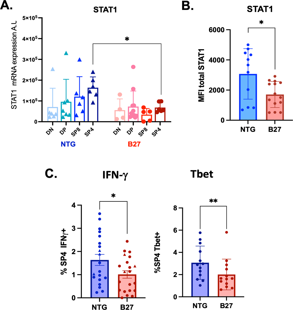
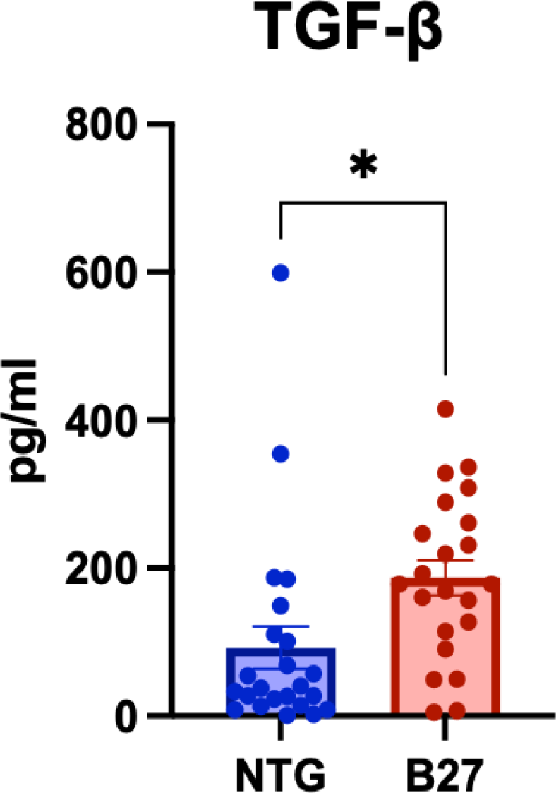
Results: Thymic architecture and thymocytes’ number were comparable between B27-rats and NTG controls. No significant differences between NTG and B27 rats were observed in STAT3 expression across thymocyte subsets or IL-17 thymic levels, indicating that the Th17 program was not intrinsically altered. In contrast, SP4 thymocytes from B27-rats exhibited a marked reduction in STAT1 expression at both mRNA and protein levels, accompanied by decreased IFN-γ + SP4 and T-bet + SP4 (Figure 1). Importantly, TGF-β levels were significantly increased in the thymus of premorbid B27-rats (Figure 2), whereas no differences were detected in the bone marrow or intestinal tissue. Despite elevated thymic TGF-β levels, the proportion of FoxP3 + SP4 was unaffected in B27-rat.
Conclusions: This study identifies early thymic dysregulation concerning the Th1/IFN/STAT1 pathway in premorbid HLA-B27 transgenic rats, associated with a thymus-specific increase in TGF-β. These alterations likely impair Th1 differentiation at the level of CD4 + T-cell direct precursors, i.e. SP4 thymocytes, which could thus favor abnormal Th17-driven responses characteristic of SpA. Our findings suggest that TGF-β could be the driver of Th1 program inhibition in B27-rat’s thymus.
Th1 program is downregulated in SP4 thymocytes from B27-rats. ( A ) STAT1 expression in DN, DP, SP8 and SP4 thymocytes from premorbid NTG and B27 rats. ( B ) Thymocytes from NTG and B27 rats were analysed by flow cytometry for cell-surface expression of TCRαβ, CD4 and CD8, and intra-cellular expression of STAT1, IFN-γ, Tbet. Data are gated on TCR〈®+CD4+CD8- thymocytes (SP4). STAT1 expression is lower in B27’s SP4 than NTG’s. ( C ) IFN-γ+SP4 and Tbet+SP4 are reduced in B27 rats.
TGF-β levels are increased in the thymus from B27-rats as compared to NTG’s.
REFERENCES: [1] Glatigny, S. et al, Arthritis Rheum 2012
[2] Fert, et al, Arthritis Rheum, 2014
[3] Araujo, L.M. et al, A&R, 2014
[4] Cherqaoui, B. et al, Frontiers Immunol., 2024
Acknowledgments: NIL.
Disclosure of Interests: None declared.