fetching data ... 
Background: Enthesitis is characterized by inflammation in and around the tendon insertion site into the bone and is key for spondyloarthritis (SpA) [1,2]. Immune cells playing a significant role in this inflammation are mainly neutrophils, macrophages and tissue resident MAIT cells, γδ T cells and innate lymphoid cells type 3. Chronic persistence of IL-17 and IL-23, produced by these cells, eventually leads to enthesophyte formation [1-3]. Currently, animal models for spondyloarthritis show enthesitis, but not always in combination with subsequent bone formation [4-6]. Besides, most models are transgenic, meaning that enthesitis develops spontaneously and is not the result of a mechanical trigger combined with immune activation as suggested to happen in SpA patients [7]. We therefore introduce a new potential mouse model of SpA enthesitis, where inflammation is mechanically induced and endochondral bone formation is observed.
Objectives: Evaluate the potential to study enthesitis and endochondral bone formation in a mechanically induced, inflammatory knee joint laxity mouse model.
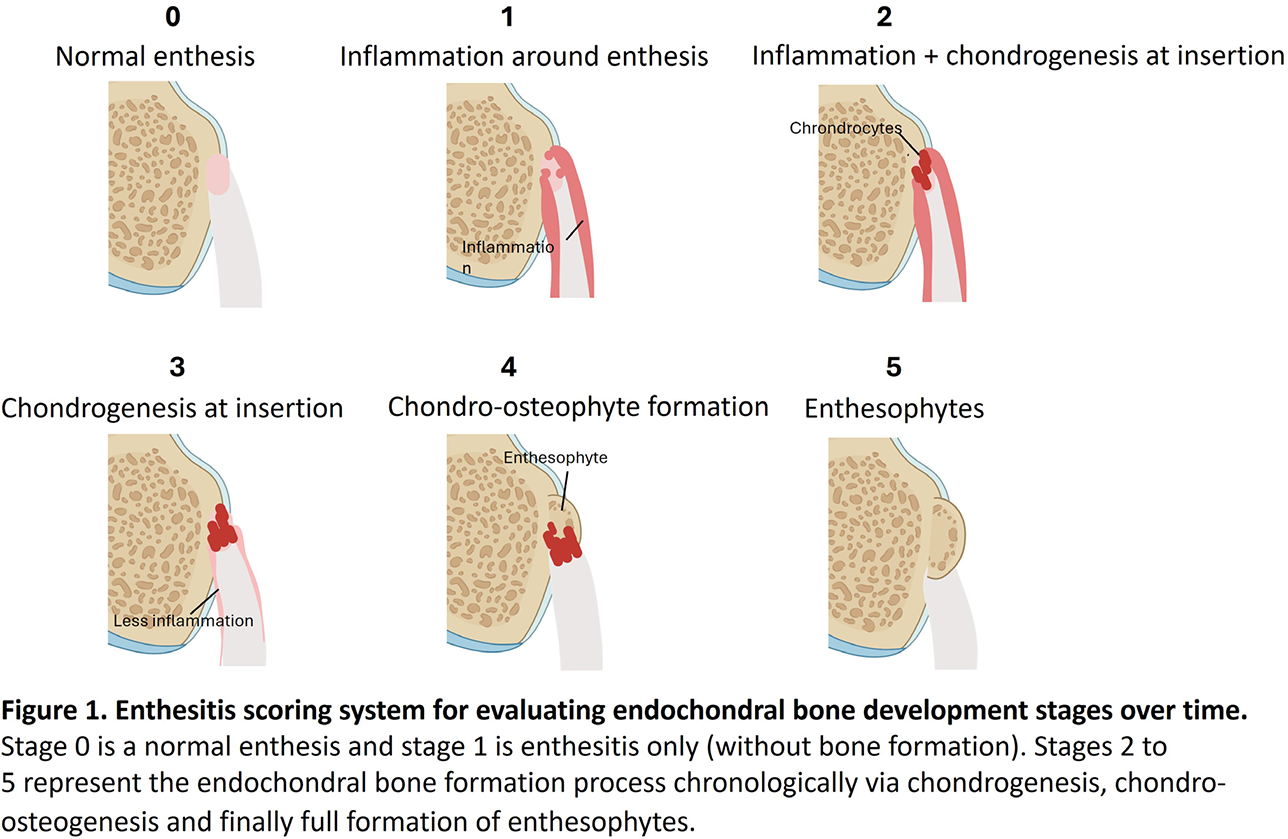
Methods: The collagenase induced osteoarthritis (CiOA) model was used in C57Bl/6 mice [8], and is known for enthesophyte formation at the collateral ligament. Collagenase was intra-articularly injected in the right knee on days 0 and 2. Mice were sacrificed on days 3, 7, 21 or 42 (CiOA time series) or days 4 or 56 (CiOA male/female study). Right knees were isolated for histological analysis of the enthesis, with H&E, Safranin O/Fast Green and anti-neutrophil antibody NIMP.R14 used for staining. We developed an enthesitis histological scoring system, based on known pathophysiological phases, with multiple stages to evaluate enthesitis features and endochondral bone development over time (Figure 1)
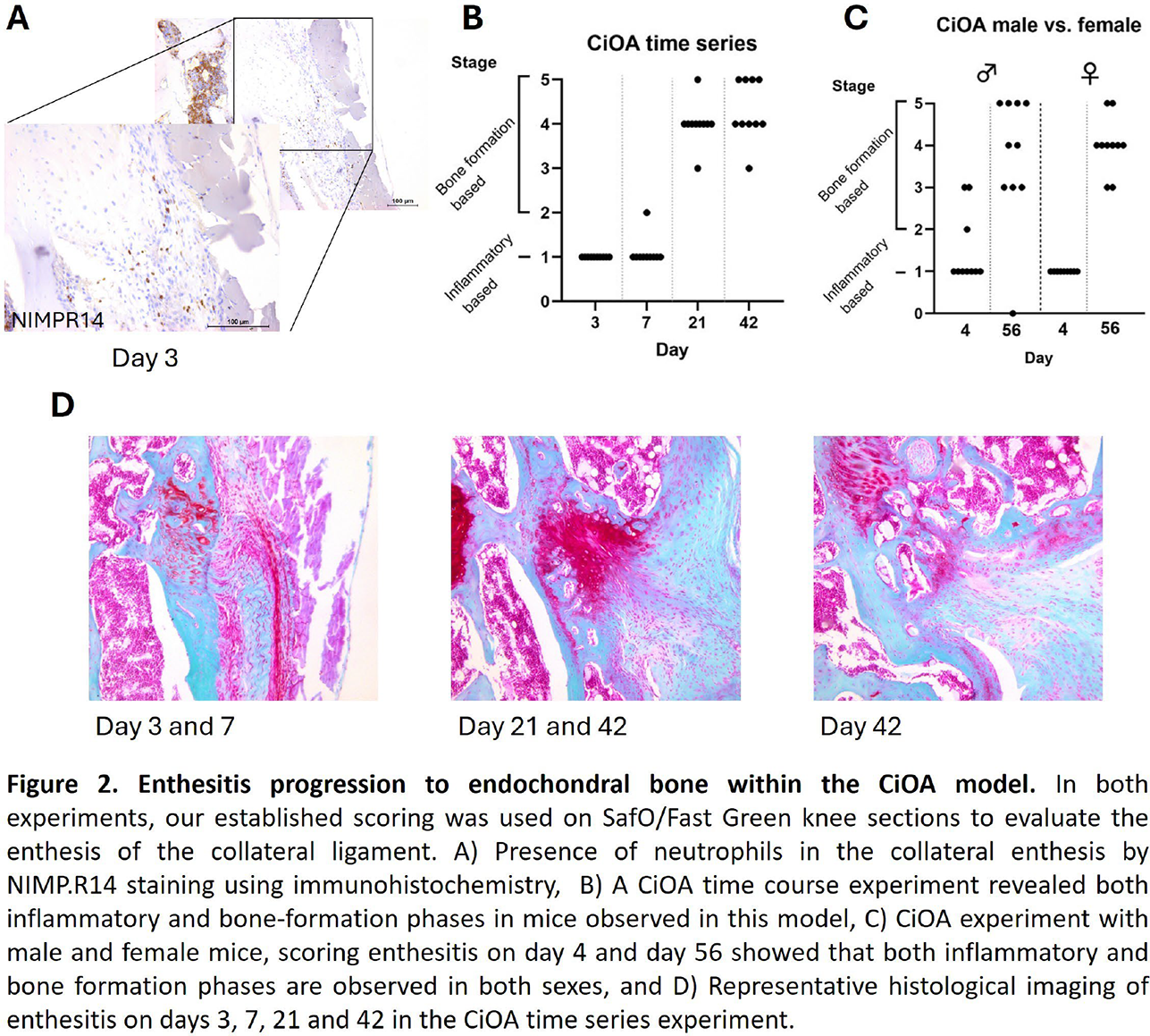
Results: Enthesitis was clearly present on days 3 and 7 of the CiOA model, as shown by neutrophils surrounding the enthesis (Figure 2). Thereafter, enthesophytes started to develop. Mice on day 21 showed predominantly chondrogenesis together with enthesophyte formation, whereas this was more spread on day 42 and fully formed enthesophytes were observed. This indicates that mice in this model progress from chondrogenesis to enthesophytes over time. To examine potential sex-differences in this novel enthesitis/SpA-like model, we compared males and females in a second experiment, focusing solely on an early inflammatory timepoint and a later bone formation timepoint. For both sexes, the same degree of enthesitis was observed on day 4 (Figure 2C). Although both timepoints are on average equal, males showed relatively more variation in this model compared to females.
Conclusions: We showed that enthesitis of the collateral ligament is present in the CiOA model which in time progresses into an enthesophyte by endochondral bone formation. These SpA features are present in both males and females and resemble to what is seen in spondyloarthritis patients, including the enthesitis onset triggered by mechanical strain combined with inflammation. Therefore, we conclude that the CiOA model and our scoring system can be used to study the mechanism of enthesitis and endochondral bone formation in spondyloarthritis.
REFERENCES: [1] van de Sande MGH, Elewaut D. Pathophysiology and immunolgical basis of axial spondyloarthritis. Best Pract Res Clin Rheumatol. 2023;37(3):101897.
[2] Schett G, Lories RJ, D’Agostino MA, Elewaut D, Kirkham B, Soriano ER, et al. Enthesitis: from pathophysiology to treatment. Nat Rev Rheumatol. 2017;13(12):731-41.
[3] Sherlock JP, Joyce-Shaikh B, Turner SP, Chao CC, Sathe M, Grein J, et al. IL-23 induces spondyloarthropathy by acting on ROR-gammat+ CD3+CD4-CD8- entheseal resident T cells. Nat Med. 2012;18(7):1069-76.
[4] Rahman MA, Thomas R. The SKG model of spondyloarthritis. Best Pract Res Clin Rheumatol. 2017;31(6):895-909.
[5] Breban M, Glatigny S, Cherqaoui B, Beaufrere M, Lauraine M, Rincheval-Arnold A, et al. Lessons on SpA pathogenesis from animal models. Semin Immunopathol. 2021;43(2):207-19. [6] Vieira-Sousa E, van Duivenvoorde LM, Fonseca JE, Lories RJ, Baeten DL. Review: animal models as a tool to dissect pivotal pathways driving spondyloarthritis. Arthritis Rheumatol. 2015;67(11):2813-27.
[7] McGonagle D, Tan AL, Benjamin M. The biomechanical link between skin and joint disease in psoriasis and psoriatic arthritis: what every dermatologist needs to know. Ann Rheum Dis. 2008;67(1):1-4.
[8] van der Kraan PM, Vitters EL, van Beuningen HM, van de Putte LB, van den Berg WB. Degenerative knee joint lesions in mice after a single intra-articular collagenase injection. A new model of osteoarthritis. J Exp Pathol (Oxford). 1990;71(1):19-31.
Acknowledgments: NIL.
Disclosure of Interests: Donna Vegt Eli Lilly, Arjen Blom: None declared, Monique Helsen: None declared, Birgitte Walgreen: None declared, Elly Vitters: None declared, Henk van Beuningen: None declared, Emmerik F.A. Leijten Eli Lilly, Novartis, Irene E. van der Horst-Bruinsma UCB, Eli Lilly, Marije Koenders Eli Lilly, Contura, Citryll, Esmeralda Blaney Davidson Eli Lilly.