fetching data ... 
Background: Rheumatoid arthritis (RA) is an autoimmune disease in which autoreactive CD4 + T cells infiltrate the joints, promoting chronic inflammation and autoantibody production. Age-associated immune dysfunction increases susceptibility to autoimmune diseases such as RA [1]. RA T cells display features of premature aging, including shortened telomeres and a pro-inflammatory cytokine profile that contributes to bone and joint destruction [2–6]. Premature T cell aging thus favors the emergence of pathogenic T cell subsets that drive disease activity and progression.
We have investigated the clonality and transcriptional profiles of distinct CD4 + T cell subpopulations from patients with RA using single-cell RNA sequencing (scRNA-seq). Based on these preliminary scRNA-seq data, CCR7 − CD27 − CD4 + T cells exhibited clonal expansion and a cytotoxic transcriptional program. The loss of CCR7 and CD27 expression was associated with the upregulation of cytotoxic effector molecules, including granzymes, perforin, KLRG1, and NKG7, as well as the migration-associated chemokine CCL5.
Objectives: The aim of this study was to investigate RA CD4 + T cells, characterized by the loss of the differentiation markers CCR7 and CD27. Functional consequences were assessed by expression analysis of cytotoxic effector molecules, including granzymes and perforin, and by functional in vitro assays of distinct T cell subsets in the context of RA.
Methods: Peripheral blood mononuclear cells (PBMCs) from RA patients (n=64) and synovial fluid (SF) samples (n=18) were isolated by density gradient centrifugation. Synovial membrane (SM) was dissected, cleared of adipose tissue, mechanically minced, and enzymatically digested (n=14). Viable cells were counted and subsequently stained for downstream analyses. Surface and intracellular T cell markers, including PD-1, CD25, ThPOK, and granzym B (GzmB), were analyzed by flow cytometry.
To assess cytotoxic T cell function, an in vitro redirected lysis assay using the P815 cell line was performed. CD4 + T cells were sorted into CD27 + and CD27 − populations by fluorescence-activated cell sorting. P815 cells were cultured under serum-free conditions, labeled with eFluor™ 670 proliferation dye, coated with α-CD3, and co-cultured with CD4 + T cells at a 1:10 ratio for 72h in the Incucyte SX5 live-cell imaging system. Target cell death was quantified by Annexin V staining.
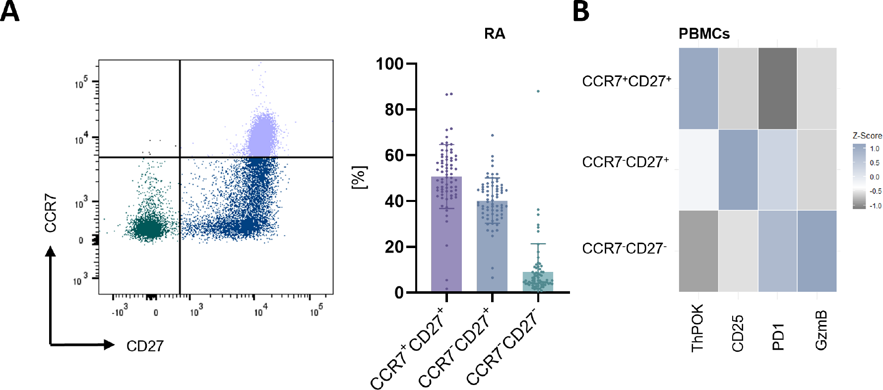
Results: Analysis of CCR7 and CD27 defined CD4 + T cell subsets in peripheral blood revealed that the majority of cells exhibited a CCR7 + CD27 + (51.28%) phenotype, followed by CCR7 − CD27 + T cells (39.78%), whereas CCR7 − CD27 − cells (9.19%) represented a minor fraction (Figure 1A). Notably, the frequency of CCR7 − CD27 − T cells correlated with clinical disease parameters, including DAS28 (n=38, *p=0.03, r=0.36), SDAI (n=29, *p=0.02, r=0.44), and C-reactive protein (CRP) (n=38, **p=0.007, r=0.43). Flow cytometric phenotyping confirmed distinct subset-specific expression profiles: CCR7 + CD27 + T cells displayed high expression of ThPOK, accompanied by low levels of PD-1, CD25, and GzmB. The loss of CCR7 was associated with downregulation of ThPOK and increased expression of CD25 and PD-1, whereas subsequent loss of CD27 resulted in marked upregulation of cytotoxic effector molecules, including GzmB, along with further increased PD-1 expression and reduced CD25 levels (Figure 1B).
CD4 + T cells from RA patients (n=3) induced efficient target cell killing, with CCR7 − CD27 − CD4 + T cells mediating more rapid cytotoxicity than CCR7 + CD27 + CD4 + T cells. Cytotoxic activity peaked within the first 30 hours and subsequently declined, whereas CCR7 + CD27 + T cells contributed relatively more to target cell lysis at later time points. The area under the curve (AUC) analysis confirmed a higher killing rate after 72 h and showed a total area of 1990 for CCR7 + CD27 + CD4 + T cells compared to 1632 for CCR7 − CD27 − CD4 + T cell killing. In comparison, T cells from healthy donors (HD) (n=2) revealed a lower killing rate (AUC of CCR7 + CD27 + CD4 + T cells = 832.6 and AUC of CCR7 − CD27 − CD4 + T cells = 1053).
Comparative analysis of SF and SM T cells demonstrated a pronounced shift in subset distribution, characterized by a marked reduction of CCR7 + CD27 + T cells (SF=8.3%; SM=5.7%) and an accumulation of CCR7 − CD27 + (SF=62.1%; SM=69.8%) and CCR7 − CD27 − (SF=28.7%; SM=23.6%) T cells. The MFI of PD-1 expression on CCR7 − CD27 − CD4 + T cells increased significantly in SF (MFI=1502) and SM (MFI=931.1) compared to blood (MFI=238.8) (****p<0.0001). In contrast, the frequency of GzmB + CCR7 − CD27 − CD4 + T cells decreased in SF and SM (blood=23.64%; SF=8.9%; SM=9.6%).
Conclusions: Our data showed that CCR7 - CD27 - CD4 + T cells from RA patients are associated with clinical RA markers, exhibit a cytotoxic phenotype and are capable of lysing target cells. These T cells contain high amounts of granzymes, which they can release faster than CD27 + CD4 + T cells. Furthermore, these cells appear to migrate into the tissue and synovial fluid but do not display the cytotoxic phenotype observed in the peripheral blood. We demonstrated that increased PD-1 expression within this cell subset is associated with downregulation of granzyme B, resulting in attenuation of cytotoxic function. Further studies are required to elucidate the functional role of these CCR7 - CD27 - CD4 + T cells within the tissue.
REFERENCES: [1] Großkopf, A., Simm, A., 2022. Alterung des Immunsystems
[2] Cope, A.P., 2008. T cells in rheumatoid arthritis
[3] Gao, Y., Cai, W., Zhou, Y., Li, Y., Cheng, J., Wei, F., 2022. Immunosenescence of T cells: a key player in rheumatoid arthritis
[4] Koetz, K., Bryl, E., Spickschen, K., O’Fallon, W.M., Goronzy, J.J., Weyand, C.M., 2000. T cell homeostasis in patients with rheumatoid arthritis
[5] Smolen, J.S., Aletaha, D., McInnes, I.B., 2016. Rheumatoid arthritis
[6] Weyand, C.M., Goronzy, J.J., 1997. PATHOGENESIS OF RHEUMATOID ARTHRITIS.
Acknowledgments: NIL.
Disclosure of Interests: None declared.