fetching data ... 
Background: Rheumatoid arthritis-associated interstitial lung disease (RA-ILD), a systemic autoimmune disorder, is characterized by joint lesions and diffuse interstitial lung alterations, and it presents a poor prognosis. The median survival duration of patients with RA-ILD is approximately 2.6-3.5 years. Recently, mesenchymal stem cells (MSCs) have demonstrated significant potential in suppressing inflammatory damage to tissues and organs, including the lungs and joints, while also promoting tissue repair and exerting anti-fibrotic effects. Consequently, MSCs are anticipated to emerge as a potential therapeutic approach for RA-ILD.
Objectives: Given that MSCs have the capacity to reduce the recruitment of inflammatory immune cells and alleviate pulmonary fibrosis, and hepatocyte growth factor (HGF) can promote the degradation of the extracellular matrix and exert an anti-inflammatory effect, MSCs modified with HGF (HGF-MSCs) can create an anti-inflammatory, anti-fibrosis, and stem-cell-colonization microenvironment. In this study, HGF-MSCs were constructed to investigate their effect on joint inflammatory injury in the RA-ILD model. Moreover, this study aims to clarify the efficacy of HGF-MSCs in reducing inflammatory injury in the lungs of the RA-ILD model, thereby laying the groundwork for the development of novel, precisely targeted stem-cell therapy drugs for RA-ILD.
Methods: We constructed a HGF overexpression vector and obtained mesenchymal stem cells (MSCs) with high HGF expression through lentiviral infection of cells. The HGF-overexpressing MSCs (HGF - MSCs) were used to rescue the mouse model of RA-ILD. Subsequently, the joint pathology (using hematoxylin and eosin (H&E), Safranine O-Fast Green, and tartrate-resistant acid phosphatase (TRAP) staining) and lung pathology (using H&E staining) of the animal model were investigated. Simultaneously, the degree of inflammatory injury in the joints and lungs was quantified through pathological analysis. Additionally, cell apoptosis in the lung tissue was detected by the terminal deoxynucleotidyl transferase dUTP nick end labeling (TUNEL) assay to evaluate the therapeutic effect of HGF-MSCs on lung injury.
HGF-MSCs alleviated joint damage in RA-ILD mouse model
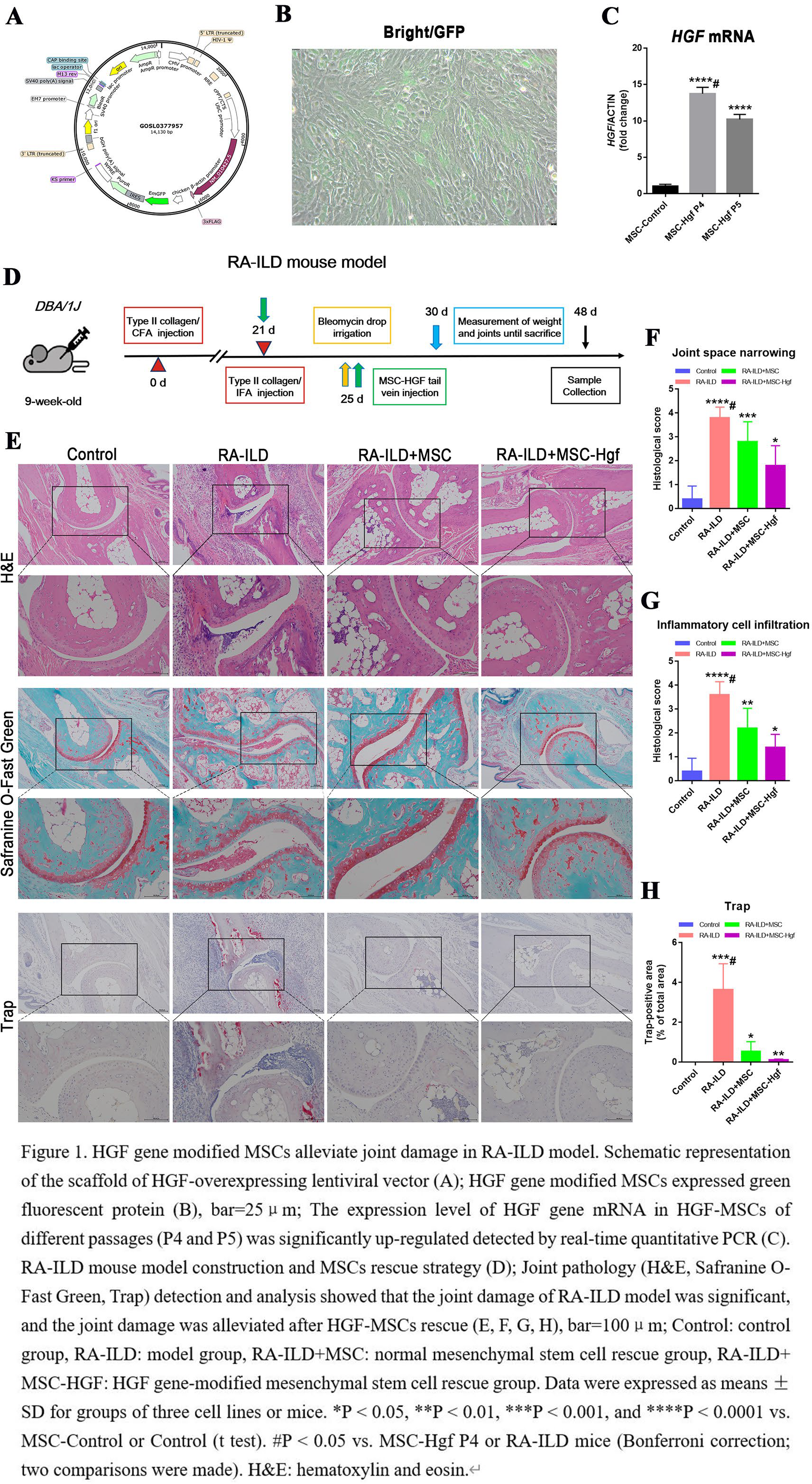
We used a lentivirus-packaged HGF overexpression vector (Figure 1 A) to infect mouse mesenchymal stem cells, thereby obtaining HGF-MSCs cells (Figure 1 B). Real-time quantitative PCR indicated that the expression level of HGF mRNA was significantly increased in the overexpressed cells (Figure 1 C). The RA-ILD model was successfully established, and a rescue strategy using HGF-MSCs was designed (Figure 1 D). Through pathological staining and analysis of the joint tissue of the model, it was found that, when compared with the control group (Control), the joint space in the RA-ILD group (RA-ILD) was narrowed, the infiltration of immune and inflammatory cells was increased, and the articular cartilage damage was obvious. In contrast, joint damage was significantly reduced after rescue with MSCs or MSCs-HGF. The MSCs-Hgf rescue group (RA-ILD+MSCs-Hgf) exhibited the least degree of injury (Figure 1 E, F, G). Moreover, TRAP staining for osteoclasts revealed that the model group (RA-ILD) had significantly more osteoclasts than the Control group (Control), whereas the MSCs rescue group (RA-ILD+MSCs, RA-ILD+ MSCs-HGF) had fewer osteoclasts (Figure 1 E, H). The aforementioned results imply that MSCs rescue (particularly MSCs-HGF) can alleviate joint pathological damage and substantially reduce bone destruction in the RA-ILD mouse model.
HGF-MSCs alleviate lung injury in RA-ILD mice
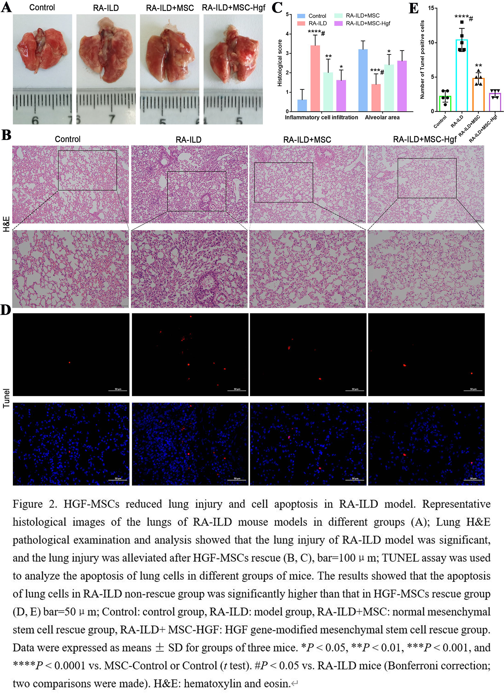
Through a comprehensive comparison of the lungs in different groups of mouse models, it was found that the lungs of RA-ILD exhibited hemorrhagic necrosis. After mesenchymal stem cells (MSCs) treatment, the hemorrhagic necrosis of the lungs in the model group was significantly reduced (Figure 2A). As demonstrated by hematoxylin and eosin (H&E) staining of lung tissues (Figure 2 B and C), when compared with the control group, the model group (RA - ILD) had a reduced alveolar area, thickened alveolar septum, and increased pulmonary interstitial inflammatory cells infiltration. After MSCs treatment, the infiltration of pulmonary interstitial inflammatory cells decreased, and pulmonary interstitial proliferation was significantly improved. The MSCs-Hgf rescue group (RA-ILD+MSCs-Hgf) showed the lowest degree of lung injury. Additionally, TUNEL was used to detect the apoptosis of cells in different groups. The results confirmed that the apoptosis of lung cells in the model group (RA - ILD) was notable, and the apoptosis of lung cells in the MSCs-Hgf rescue group (RA-ILD+MSCs-Hgf) was significantly reduced (Figure 2 D, E). These results confirmed that MSCs-Hgf rescue could inhibit the infiltration of inflammatory immune cells and reduce the apoptosis of lung cells, thereby alleviating the inflammatory injury of the RA-ILD lung.
Conclusions: Our research group has successfully constructed HGF-modified MSCs and a mouse model of RA-ILD. Subsequently, we used HGF-MSCs to treat the RA-ILD mouse model. It was found that HGF-MSCs can notably alleviate the pathological damage to joints and lungs, suppress the infiltration of immune inflammatory cells, and decrease cell apoptosis in the RA-ILD mouse model. These findings provide a basis for the subsequent development of precise stem cells therapy for RA-ILD.
REFERENCES: NIL.
Acknowledgments: NIL.
Disclosure of Interests: None declared.