fetching data ... 
Background: Despite advances in induction and maintenance therapies, relapse of antineutrophil cytoplasmic antibody–associated vasculitis (AAV) remains a major unsolved clinical challenge. Although interferon (IFN) signaling pathways have been implicated in AAV pathogenesis, most transcriptomic studies have been cross-sectional or focused on baseline disease activity. How early post-treatment changes in gene expression relate to subsequent relapse risk remains poorly understood.
Objectives: To investigate early post-treatment changes in peripheral blood gene expression profiles in patients with AAV and to identify molecular pathways and immune cell dynamics associated with relapse, with a particular focus on interferon signaling.
Methods: We performed bulk RNA sequencing of peripheral blood samples from 22 patients with newly diagnosed AAV, including 7 patients with granulomatosis with polyangiitis and 15 with microscopic polyangiitis. Samples were collected before treatment initiation and at 4 weeks after induction therapy (44 samples). Relapse was defined as clinical deterioration requiring modification or escalation of immunosuppressive therapy during follow-up. Patients were classified into a relapse group (n=12) and a non-relapse group (n=10). Paired differential expression analysis was conducted to identify treatment-induced differentially expressed genes (DEGs) within each group. To extract relapse-associated transcriptional programs, paired pre- and post-treatment differences were calculated within each group, and these differences were subsequently compared between groups using gene set enrichment analysis (GSEA). Peripheral immune cell composition was computationally inferred using CIBERSORTx. Interferon signature scores were calculated, and correlations between immune cell changes and interferon responses were evaluated using Spearman’s correlation analysis.
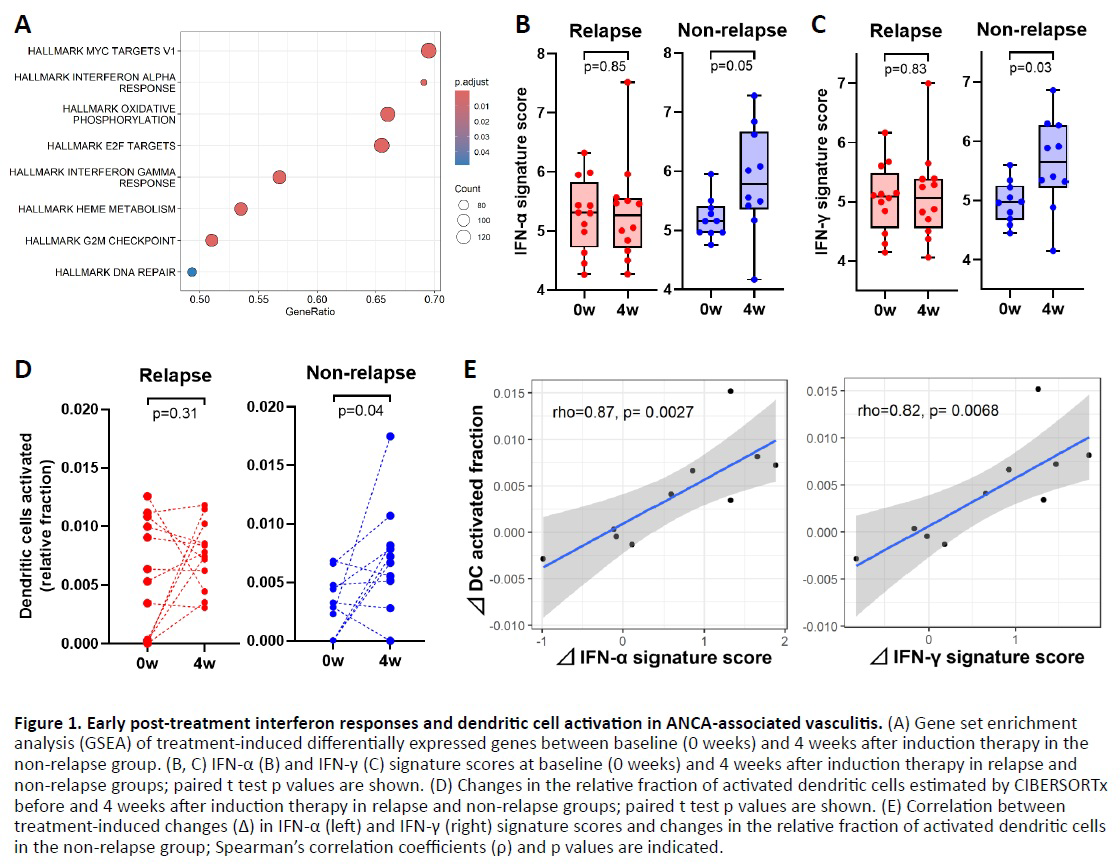
Results: Baseline demographic and clinical characteristics, including age, sex, disease subtype, ANCA specificity, and disease activity, were comparable between the two groups, except for the number of rituximab doses administered during induction therapy, which was higher in the non-relapse group (p=0.019). The magnitude and breadth of treatment-induced gene expression changes were markedly different between groups. A total of 116 DEGs were identified in the relapse group, whereas 329 DEGs were detected in the non-relapse group, indicating broader post-treatment transcriptional changes in patients who achieved sustained remission. Difference-of-differences GSEA revealed significant enrichment of interferon-related pathways in the non-relapse group, including IFN-α signaling (p=2.48×10 −7 ) and IFN-γ signaling (p=9.17×10 −8 ) (Figure 1A), whereas these pathways were not enriched in the relapse group. IFN-α and IFN-γ signature scores increased significantly after treatment in the non-relapse group, whereas no significant changes were observed in the relapse group (Figure 1B, 1C). CIBERSORTx analysis demonstrated a significant increase in activated dendritic cells (DCs) at week 4 after treatment in the non-relapse group (p=0.044), while no significant changes were observed in the relapse group (Figure 1D). In the non-relapse group, changes in activated DC fractions showed strong positive correlations with increases in IFN-α and IFN-γ signature scores (ρ=0.87 and 0.82, respectively) (Figure 1E). In contrast, these correlations were absent in the relapse group.
Conclusions: In this study, patients who achieved sustained remission showed broader treatment-induced gene expression changes, enrichment of IFN-α and IFN-γ signaling pathways, and a significant increase in activated dendritic cells early after induction therapy. Notably, interferon signature upregulation was strongly correlated with dendritic cell activation only in non-relapsing patients. These findings extend previous longitudinal transcriptomic studies demonstrating that early treatment-induced changes, rather than baseline expression levels, are predictive of long-term outcomes in AAV [1], and contrast with recent single-cell analyses identifying IFN-γ–driven neutrophil programs associated with relapse susceptibility [2].
Together, our results suggest that relapse risk in AAV is influenced not simply by the presence of interferon signaling, but by its cellular context, with dendritic cell–associated interferon responses reflecting a coordinated post-treatment immune state linked to sustained remission.
REFERENCES: [1] Ishizu A, et al. Prediction of response to treatment by gene expression profiling of peripheral blood in patients with microscopic polyangiitis. PLoS One, 2013, 8(5), e63182.
[2] Nishide M, et al.Neutrophil single-cell analysis identifies a type II interferon-related subset for predicting relapse of autoimmune small vessel vasculitis. Nat Commun. 2025;16(1):3581.
Acknowledgments: NIL.
Disclosure of Interests: Shuji Sumitomo Asahi-Kasei Pharma, GSK, AstaraZeneca, Abbvie, Pfizer, Eli Lilly, Chugai Pharmaceutical, Astellas, BMS, Eizai, Tanabe Pharma, Hideki Oka: None declared, Takeshi Iwasaki: None declared, Koichiro Ohmura Eisai, Gilead Sciences, AstraZeneca, GSK, Asahi-Kasei Pharma, Chugai Pharmaceutical, Tanabe Pharma.