fetching data ... 
Background: Leukocyte immunoglobulin-like receptors (LILRs) are immunomodulatory proteins predominantly expressed on immune cells. The LILR family consists of 13 members, including activating (LILRA) and inhibitory (LILRB) receptors, of which LILRA3 is the only soluble member. A naturally occurring deletion in the LILRA3 gene, resulting in a null allele and complete absence of the protein, has been associated with several immune-mediated diseases. Despite these associations, the biological function and the ligand of LILRA3 remain poorly defined. Previous studies suggest that B cells and monocytes represent primary targets of LILRA3 binding.
Objectives: In this study, we investigated the effects of LILRA3 on human B cells, an essential part of the immune system and a crucial factor in several autoimmune diseases.
Methods: Peripheral blood mononuclear cells (PBMCs) from healthy donors (n = 17) as well as patients with Systemic Lupus Erythematosus (SLE) (n = 8) and Common Variable Immunodeficiency (CVID) (n = 7) were stimulated with B cell activators including CpG (TLR-9 agonist), anti-CD40 and anti-IgM, as well as LILRA3 either alone or in combination with the respective B cell activators. The human recombinant LILRA3 used in this study was generated in a eukaryotic expression system. Proliferation of total B cells and B cell subpopulations was assessed by PKH26-dilution by flow cytometry after 5 days of incubation. Additionally, cytokine production was analyzed by intracellular immunolabeling for tumor necrosis factor (TNF) (n = 17) and Lymphotoxin-alpha (LT-α) (n = 15). To assess transcriptional changes, RNA was isolated from stimulated and unstimulated B cells (n = 4) and RNA bulk sequencing was performed.
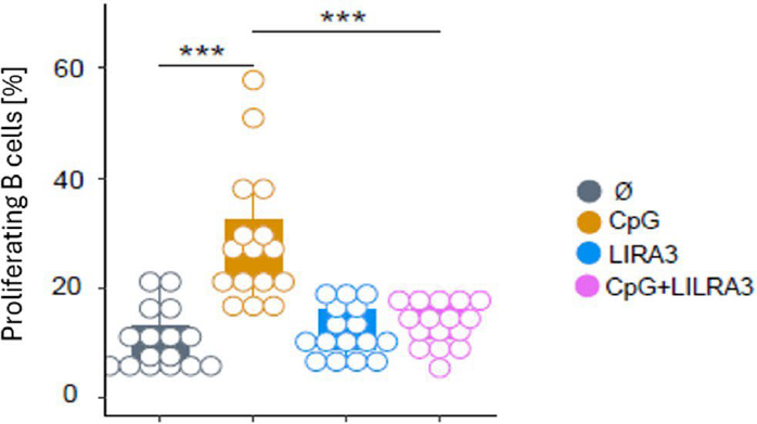
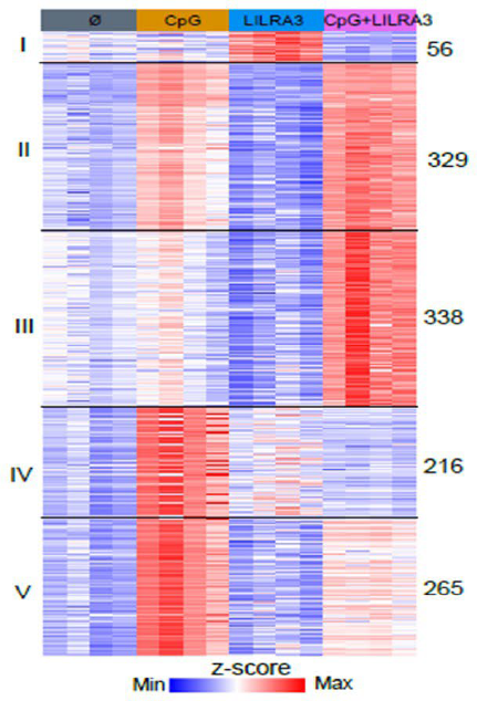
Results: Upon stimulation with LILRA3 alone no effects were detected on the level of B cells, whereas stimulation with CpG alone induced B cell proliferation. Co-stimulation of PBMCs with CpG and LILRA3 significantly reduced the B cell proliferation (p < 0.001), indicating an inhibitory effect of LILRA3 on CpG-induced B cell proliferation. This effect was consistent across all analyzed B cell subsets. A similar, though not statistically significant, inhibitory effect was observed upon co-stimulation with anti-CD40 and LILRA3. In contrast, anti-IgM-mediated B cell proliferation was not affected by co-stimulation with LILRA3. Unlike proliferation, LILRA3 does not alter CpG-induced production of pro-inflammatory cytokines such as TNF and LT- α. Similar to healthy donors, B cells from SLE and CVID patients also showed inhibitory modulation of TLR9-driven proliferation by LILRA3. RNA sequencing revealed the induction of several immunomodulatory genes, including Interleukin 10 (IL-10 ), Transforming growth factor ß (TGF-ß ) and NUAK Family Kinase 2 (NUAK2 ), as well as the suppression of pro-inflammatory pathways upon LILRA3 and CpG co-stimulation.
Conclusions: LILRA3 acts as an inhibitory modulator of B cell responses by inhibiting TLR-9 and CD40-driven proliferation. The inhibitory effect of LILRA3 comprises downmodulation of several pro-inflammatory pathways. These includes inflammatory responses and response to cytokines by promoting subcellular organelle reorganization and autophagy without affecting pro-inflammatory cytokine production. These findings provide new insight into the immunoregulatory role of LILRA3, which has previously been described as an immune-activating member of the LILR family. TLR-9 plays an important role in the pathogenesis of diseases such as SLE, making the LILRA3-mediated inhibition of TLR-9-induced B cell proliferation an interesting target for therapeutic modulation of autoimmune responses.
Stimulation of PBMCs with CpG results in enhanced proliferation of B cells, whereas stimulation with human recombinant LILRA3 protein did not alter proliferation. CpG-induced proliferation was abrogated when PBMCs were co-stimulated with CpG and LILRA3.
RNA sequencing revealed that LILRA3 co-stimulation leads to the induction of several immunomodulatory genes, such as TGF-ß and NUAK2 , as well as the suppression of pro-inflammatory pathways.
REFERENCES: NIL.
Acknowledgments: NIL.
Disclosure of Interests: Julia Grupa: None declared, Felix Mulenge: None declared, Christine Ehlers: None declared, Katja Kniesch: None declared, Bibiana Costa: None declared, Ulrich Kalinke: None declared, Torsten Witte AbbVie, Alexion, AMGEN, BMS, Celltrion, Chugai, CSL Behring, Galapagos/Alfasigma, GSK, Janssen, Lilly, Medac, Nordic, Novartis, Octapharma, Pfizer, Roche Pharma, Sanofi, Takeda, UCB, AbbVie, Indero, J&J, Novartis, UCB, AbbVie, Novartis, Takeda, Theresa Graalmann: None declared, Gerrit Ahrenstorf Alfasigma, Gilead Sciences, GSK, UCB, Johnson&Johnson, Takeda, ViiV Healthcare, Alfasigma, Gilead Sciences, GSK, Johnson&Johnson, ViiV Healthcare.