fetching data ... 
Background: Lupus nephritis (LN) is one of the most common complications of systemic lupus erythematosus (SLE). Once it progresses to end-stage renal disease (ESRD), it is associated with high mortality and is a life-threatening condition. Monocyte/macrophage (MO/MΦ) plays a crucial regulatory role in the pathogenesis of LN and is a key cell type mediating renal tissue injury. Our previous study found that activation of the kidney-derived transcription factor SIX1 is closely linked to MΦ infiltration, glycolytic pathway activation, and heightened immune inflammation, suggesting that SIX1 may be involved in inflammatory and metabolic pathways in LN kidneys.
Objectives: This study is founded upon three key aspects: (1) metabolic reprogramming in activated mononuclear–macrophages within the immune microenvironment; (2) hypoxia-induced enhancement of the glycolytic pathway; and (3) the role of Six1 as a critical regulator of hypoxic glycolysis. The objectives are to: (1) determine whether Six1 is activated and involved in renal inflammatory injury in LN patients; (2) clarify how hypoxic glycolysis promotes the inflammatory phenotype of macrophages (MΦ); and (3) elucidate the molecular mechanism by which Six1 activation regulates MΦ glycolysis, thereby identifying a potential Six1-targeted intervention to mitigate renal inflammation.
Methods: We successfully established the MRL/lpr mouse model. Kidney damage was assessed at multiple disease stages (13, 17, and 21 weeks of age) using macroscopic phenotypic evaluation, biochemical analysis, and pathological examination. Concurrently, we measured expression levels of the key molecule Six1, metabolic molecules, and inflammatory markers via molecular biology techniques. Mathematical correlation analysis was performed to evaluate the relationship between Six1 expression and glycolysis-related as well as inflammation-associated molecules. In addition, in vitro macrophage activation models from different sources were established to analyze Six1 expression and its association with metabolic and inflammatory molecules in MΦ, and to investigate the impact of Six1 overexpression on glycolytic pathway molecules.
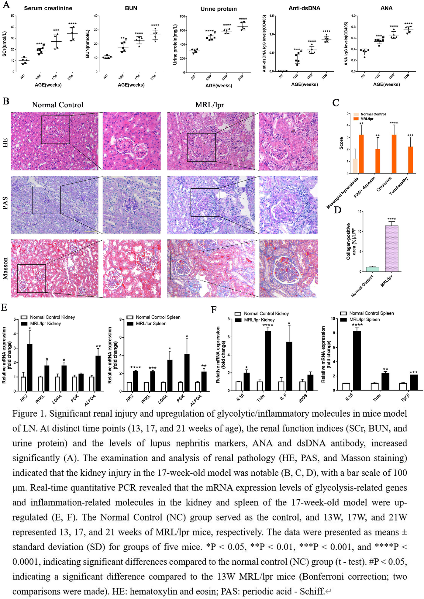
The expression of glycolysis/inflammation-related molecules was significantly upregulated in the LN mice model
Our findings confirm that in the MRL/lpr LN model, SCr, BUN, urinary protein, and LN markers ANA and anti-dsDNA antibody levels increased significantly with disease progression at 13, 17, and 21 weeks of age (Figure 1A). Renal histopathological analyses (HE, PAS, Masson staining) showed that at 17 weeks, glomerular immune complex deposition, interstitial inflammatory cell infiltration, and crescent formation were markedly elevated in LN mice compared to controls (Figure 1B). Quantitative pathological assessment and collagen deposition analysis further revealed significantly higher scores in the LN group (Figures 1C, D). qPCR results demonstrated that at 17 weeks, mRNA expression of glycolytic genes—including HK2, PFKL, ALDOA, Pgk, and Ldha—was significantly upregulated in the kidneys and spleens of LN mice versus controls (Figure 2E). Additionally, renal and splenic expression levels of inflammatory molecules such as IL-1β and TNFα were also significantly elevated in LN mice (Figure 2F). These results indicate that in the LN mouse model, both kidney and spleen exhibit metabolic reprogramming characterized by enhanced glycolysis and inflammation, concurrent with significant renal injury.
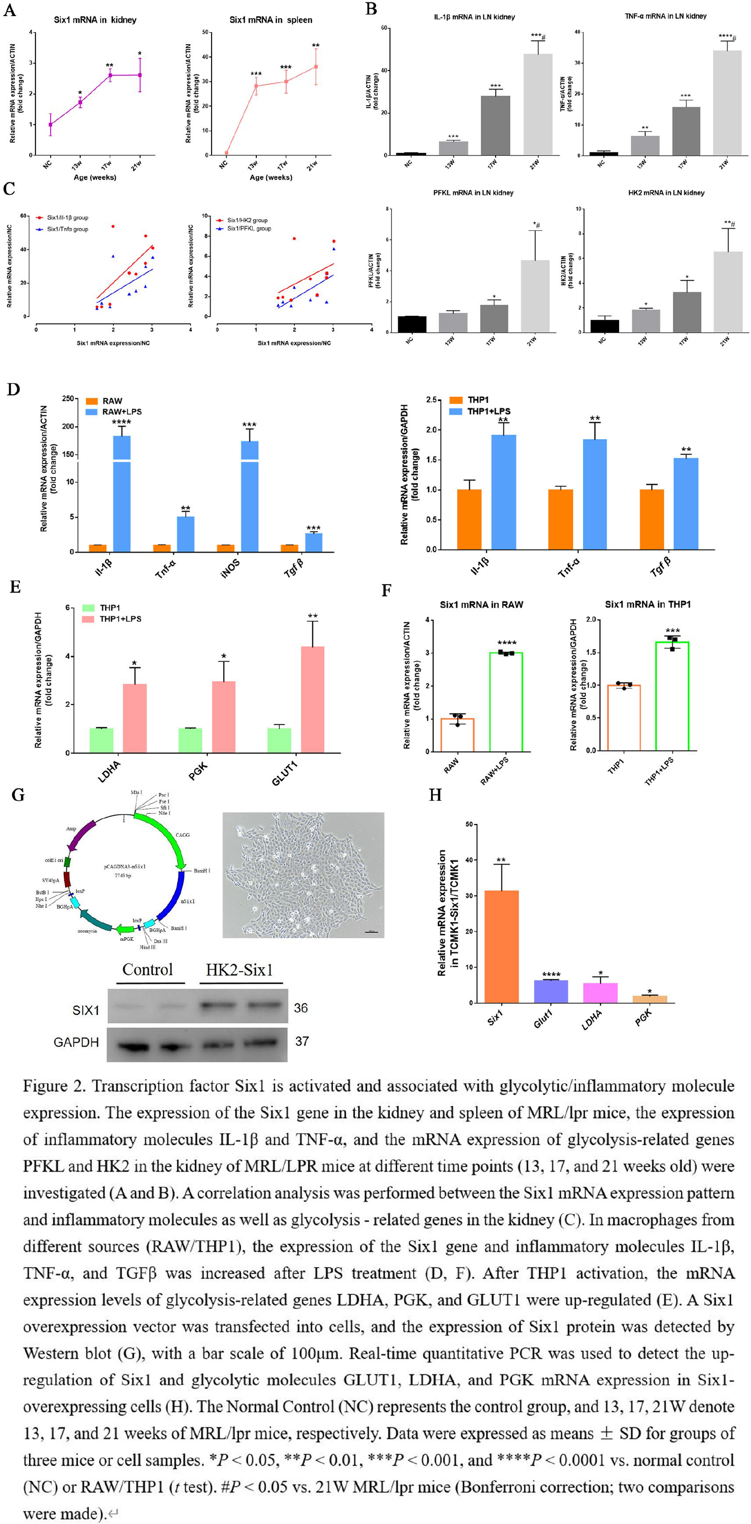
Six1 activation mediates macrophage glycolytic pathway and the release of inflammatory molecules
qPCR was used to detect Six1 gene expression in the kidneys and spleens of MRL/lpr mice. Six1 mRNA levels were significantly upregulated in both organs, with increasing expression as disease progressed (Figure 2A). Based on Six1 expression patterns and inflammatory and glycolytic gene expression in the kidneys (Figure 2B), linear correlation analysis showed that Six1 expression positively correlated with key glycolysis- and inflammation-related genes (PFKL, HK2, IL-1β, Tnfα) during disease progression (Figure 2C). In activated mouse and human macrophages in vitro, Six1 upregulation (Figure 2F) coincided with increased expression of metabolic and inflammatory markers (Glut1, Pgk, Ldha, IL-1β, Tnfα) (Figure 2D, E). To further investigate, a Six1 overexpression vector was transfected into cells to establish stable Six1-overexpressing lines (Figure 2G). Molecular assays revealed that Six1 overexpression significantly enhanced glycolysis-related gene expression (Figure 2H). These results suggest that Six1 activation in MΦ in LN promotes glycolytic pathway activation and inflammatory mediator release, contributing to renal inflammatory injury.
Conclusions: In the established MRL/lpr mouse model, Six1 expression was significantly upregulated in the kidneys and spleens and positively correlated with metabolic molecules (Pgk, Ldha) and inflammatory markers (IL-1β, Tnfα). In vitro, Six1 is highly expressed in activated monocyte–macrophages, where metabolism- and inflammation-related molecules are also upregulated. Six1 overexpression increases glycolysis-related molecule expression, suggesting its role in promoting glycolytic pathway activation and exacerbating renal inflammatory injury in LN. Mechanistically, Six1 enhances glycolytic signaling and drives metabolic reprogramming.
REFERENCES: NIL.
Acknowledgments: NIL.
Disclosure of Interests: None declared.