fetching data ... 
Background: Systemic Lupus Erythematosus (SLE) is a chronic autoimmune disease characterized by widespread inflammation and tissue damage. Lupus Nephritis (LN) represents one of the most severe complications, affecting up to 60% of patients. Despite advances in combined glucocorticoid and immunosuppressive therapies, a proportion of patients fail to achieve remission and progressively develop renal fibrosis. This distinct pathological process, driven by extracellular matrix accumulation, inevitably leads to End-Stage Renal Disease. However, the lack of approved specific anti-fibrotic therapies for LN constitutes an urgent and unmet clinical need. Pirfenidone (PFD) is a small-molecule agent with established anti-fibrotic activity, currently serving as a standard-of-care therapy for Interstitial Lung Disease. Although its therapeutic efficacy has been validated in general models of renal fibrosis, its specific application in LN remains experimentally unverified. However, whether PFD can effectively attenuate renal fibrosis specifically driven by LN, and the underlying molecular mechanisms, remain to be elucidated.
Objectives: The aim of this study is to evaluate the feasibility and effectiveness of PFD in attenuating renal fibrosis in the MRL/lpr mouse model, given its established efficacy in other renal pathologies. To elucidate the underlying molecular mechanism, this study utilized transcriptomic profiling to screen for potential therapeutic targets, leading to the identification and specific investigation of Matrix-remodeling associated 7 (MXRA7). Consequently, the role of the PFD-MXRA7 axis in mediating renal protection was validated.
Methods: (1) MRL/lpr mice were randomized into Model (vehicle) and PFD (300 mg/kg/d, oral gavage) groups from 8 to 18 weeks of age, while MRL/MpJ mice served as the Control group. (2) At the endpoint, organ indices of spleen and lymph nodes were calculated; urine and serum samples were collected to evaluate renal function (Urinary Protein/Creatinine Ratio, Urinary Microalbumin/Creatinine Ratio, Scr, Urea) and liver safety (ALT, AST). (3) Renal and pulmonary tissues were harvested for Masson and PASM staining to evaluate pathological changes. (4) RNA-seq and KEGG pathway analysis were performed on kidney tissues to screen potential targets. (5) The expression of target gene MXRA7, COL1A1 and ACTA2 was validated by RT-qPCR and Western Blot.
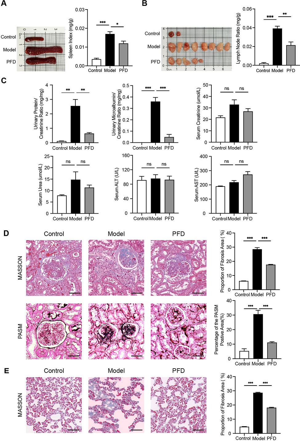
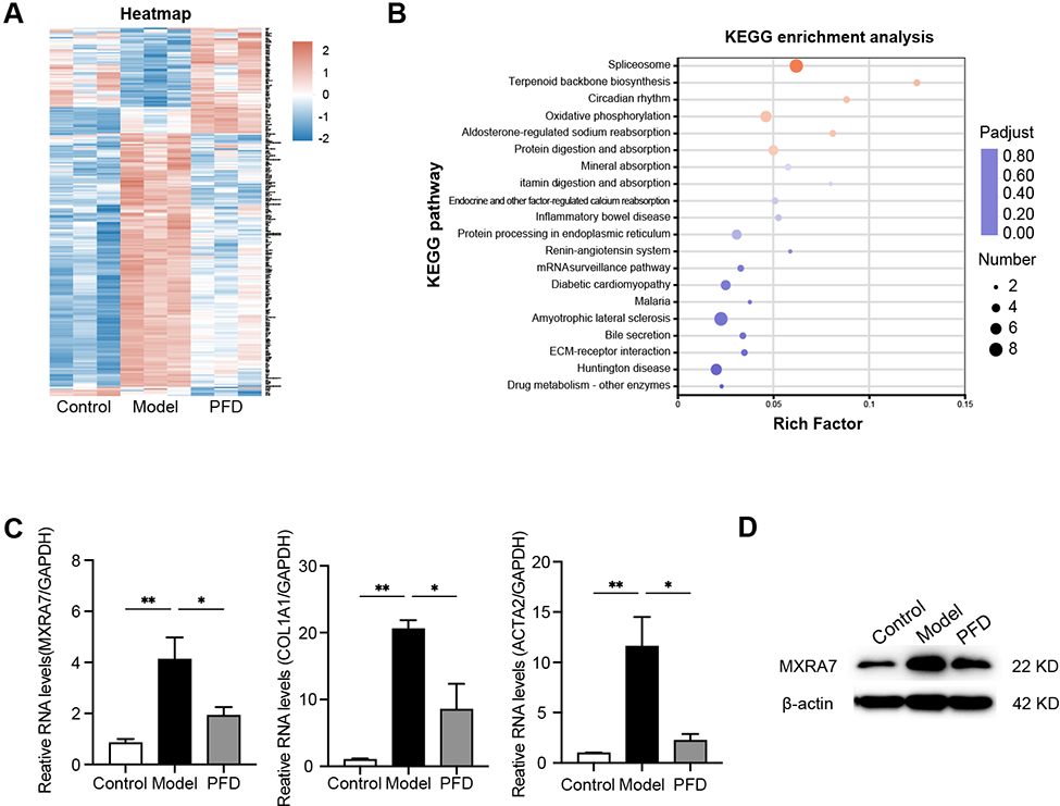
Results: PFD Ameliorates Systemic Inflammation and Mitigates Renal Fibrosis and Dysfunction in MRL/lpr Mice. The Model group exhibited severe systemic inflammation and renal dysfunction compared with the Control group. PFD treatment significantly attenuated splenomegaly and lymphadenopathy, evidenced by reduced organ indices (Figure 1A-B). Functionally, PFD preserved glomerular integrity, resulting in marked reductions in Urinary Protein/Creatinine Ratio and Urinary Microalbumin/Creatinine Ratio compared with the Model group (Figure 1C). Notably, PFD showed a favorable safety profile; serum Scr, Urea, ALT, and AST levels showed no pathological elevation and were comparable to the Control group (Figure 1C). Histologically, PFD treatment significantly ameliorated renal interstitial fibrosis and glomerular sclerosis, quantified by the reduced fibrotic and PASM-positive areas (Figure 1D). Consistent anti-fibrotic effects were also observed in lung tissues with a decrease in fibrosis area (Figure 1E), confirming the systemic efficacy of PFD. Transcriptomic Profiling Identifies MXRA7 as a Key Target of PFD-Mediated Anti-Fibrotic Effects. Transcriptomic analysis revealed distinct gene expression signatures among groups. Hierarchical clustering of DEGs (|log2FC| > 1.2) illustrated the separation between groups (Figure 2A). KEGG pathway enrichment analysis indicated that PFD treatment significantly modulated critical metabolic and structural pathways, notably “Oxidative phosphorylation”, “Aldosterone-regulated sodium reabsorption”, and “ECM-receptor interaction” (Figure 2B). Among the downregulated genes, MXRA7 was identified as a top candidate. Validation by RT-qPCR confirmed that PFD significantly reversed the aberrant upregulation of MXRA7 mRNA observed in the Model group, paralleling the reduction of key fibrotic markers COL1A1 and ACTA2 (Figure 2C). Consistent with these findings, Western Blot analysis demonstrated that PFD effectively suppressed MXRA7 protein abundance compared with the Model group (Figure 2D).
Conclusions: PFD treatment can successfully alleviate the progression of LN in MRL/lpr mice. Oral administration of PFD can reduce spleen and lymph node indices, proteinuria levels, and the degree of renal fibrosis and pathological damage. Mechanistically, PFD exerts its effects by downregulating the expression of the target gene MXRA7. PFD targeting the MXRA7 axis may become an effective drug for the treatment of renal fibrosis in LN.
Therapeutic efficacy of PFD on LN and systemic symptoms in MRL/lpr mice. (A) Gross morphology of spleens and comparison of spleen index among groups; (B) Gross morphology of lymph nodes and comparison of lymph node ratio; (C) Analysis of renal function markers (Urinary Protein/Creatinine Ratio, Urinary Microalbumin/Creatinine Ratio, Serum Creatinine, Serum Urea) and liver function enzymes (ALT, AST);(D) Renal histopathological changes and quantitative analysis of Masson and PASM staining (Scale bar = 50 μm); (E) Pulmonary histopathological changes and quantitative analysis of Masson staining (Scale bar = 50μm). *p<0.05, **p<0.01, ***p<0.001.
Screening and validation of the PFD target gene MXRA7. (A) Heatmap visualization of differentially expressed genes among groups; (B) KEGG pathway enrichment analysis of genes reversed by PFD treatment; (C) Relative mRNA expression levels of MXRA7, COL1A1 and ACTA2 detected by RT-qPCR; (D) Protein expression levels of MXRA7 determined by Western blot analysis. Data are expressed as mean±SEM(n=3). * p <0.05, ** p <0.01.
REFERENCES: NIL.
Acknowledgments: NIL.
Disclosure of Interests: None declared.