fetching data ... 
Background: Lupus nephritis is a common, life-threatening complication of systemic lupus erythematosus in children that frequently does not respond to conventional therapies. Identification of novel targeted therapies to treat lupus nephritis requires a more granular understanding of the autoimmune response. Previous spatial -omics methods have been limited to 1000 or fewer analyzed genes, limiting the identification of cell subtypes, particularly in immune cells. Furthermore, the measurement and analysis of RNA transcripts alone provides an incomplete picture of cellular function, which is primarily driven by the expression and action of proteins.
Objectives: The purpose of this study was to leverage a high-plex, integrated subcellular spatial transcriptomics and proteomics platform to map 6,000 genes and 64 proteins across hundreds of thousands of cells from pediatric cLN biopsies, control kidney samples (nephrectomy specimens from patients with a non-inflammatory condition), and tonsils. By comparing diseased kidneys to tonsillar immune architecture, we aim to identify aberrant lymphocyte developmental states and immune circuits that may represent novel therapeutic targets. By studying stromal cellular stress responses and interactions patterns with immune cells, particularly T cells, we intend to identify putative stromal cell targets of the autoimmune response. Finally, we hope to use the protein data to better define normal and aberrant immune cell subsets and their functions.
Methods: Three patients with biopsy-confirmed class III or IV cLN, and a patient who received a nephrectomy for a perihilar mass, were enrolled in a no-contact clinical study approved by the Seattle Children’s Hospital Institutional Review Board (IRB). Subsequently, previous biopsies obtained for clinical diagnostic purposes at or near diagnosis were subject to the CosMx Human Multiomics platform (Bruker). De-identified tonsillar tissue obtained as part of routine clinical care was subject to the same platform. Cell profiles were subject to Insitutype , a Bayesian method for identifying cell types, using the Kidney Precision Medicine Project (KPMP) v2 reference cell types [1]; and subject to unsupervised clustering via principal component analysis (PCA) and uniform manifold approximation and projection (UMAP) using the transcriptional data only. Differential gene testing and spatial relationship analyses were performed for lymphocyte cell subclusters that were shared between tonsillar and cLN tissues. The following analyses are ongoing as of submission of this abstract: differentially expressed gene analyses, cell interaction network analyses, and single cell morphometric analysis of the subcellular resolution protein data.
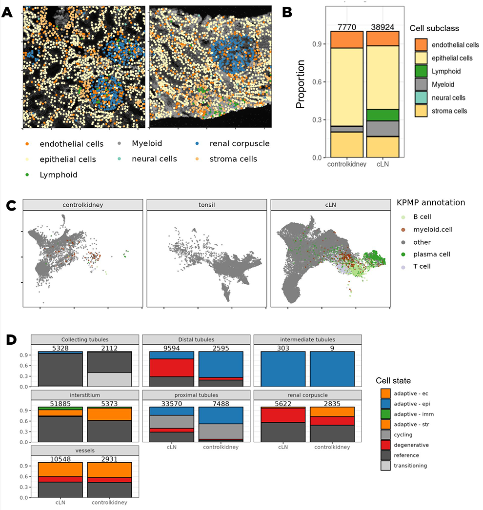
Results: Supervised cell identification of 47,470 kidney cells revealed expected spatial relationships between cells in the kidney (Figure 1A). A large increase in the number of lymphocytes observed in cLN (9.2% of all cells profiled), compared to the non-inflammatory kidney control (0.5% of all cells profiled; Figure 1B). Approximately 33% of these highly abundant lupus nephritis lymphocytes were mapped to clusters also observed in the healthy tonsillar tissue in PCA space (Figure 1C). Examination of KPMP reference-defined cell states revealed that patients with lupus nephritis exhibit significant stromal degeneration signatures in the distal convoluted tubules, compared to the nephrectomy control. Both the nephrectomy control and lupus samples exhibited degenerated cell states in blood vessel cell types, which may represent hypoxic injury related to tissue collection (Figure 1D).
Conclusions: The transcriptional overlap between tonsillar and lupus nephritis lymphocytes suggests that there is aberrant lymph-node like differentiation of B and T cells within the kidney in patients with cLN. This is congruent with past reports of tertiary lymphoid structures being observed in autoimmune diseases. This in situ autoreactive lymphocyte development may be a keystone in the pathogenesis of lupus nephritis, as evidenced by the recent success of deep CD19-directed B cell-depleting therapies. The high proportion of distal tubular cells that have degeneration signatures may be indicative of a hypoxic environment downstream of the glomeruli, or related to the spatial proximity of these cells with the glomerulus; further spatial analyses using this dataset will help determine which of these is playing a role.
Analysis of spatial transcriptomics data. (A) Spatial plot showing cells annotated by Kidney Precision Medicine Partnership (KPMP)-defined cell subclasses using the transcriptomics data. Behind each plot is a grayscale image of fluorescnce from a PanCK marker, which was used for cell segmentation. (B) Proportion of cell subclasses in control kidney vs chilhood lupus nephritis (cLN). (C) Uniform manifold projection and approximation (UMAP) plot of control kidney, tonsils, and cLN. (D) Proportions of KPMP-defined cell states across nephron compartments in control kidneys and cLN. ec = epithelial cell; epi = epithelial cells; str = stromal cells; imm = immune cell.
REFERENCES: [1] Lake, B. B., Melo Ferreira, R., Hansen, J., Menon, R., Basta, J., Thiessen Philbrook, H., Reinert, S., Fallegger, R., Lagwankar, A. K., Chen, X., Maity, S., Djambazova, K. v., Gorman, B. L., Lucarelli, N., Gisch, D. L., Schmidt, I. M., Nair, V., Alakwaa, F., Kefaloyianni, E., … Jain, S. (2025).
Cellular and Spatial Drivers of Unresolved Injury and Functional Decline in the Human Kidney
.
Acknowledgments: NIL.
Disclosure of Interests: Nicholas Hasle N.H. and his spouse hold shares in Immunome, Inc. They previously held shareholders in Bristol-Myers Squib., Robyn C. Reed: None declared, Ginny Schultz: None declared, Mark Conner: None declared, Rebecca Martin R.M. holds stock in Bristol Meyer Squibb & Merck., Shaun W. Jackson S.W.J. is a consultant for Amgen, Bristol-Myers Squib, Merck, and Palleon Pharmaceuticals. He previously served as a consultant for IgM BioSciences and Sail BioMedicines.