fetching data ... 
Background: Evidences show that antimalarial agents of artemisinin and its derivatives such as artesunate may inhibit proinflammatory cytokines secretion from human rheumatoid arthritis (RA) fibroblast-like synoviocytes (FLS) in vitro. It has also been demonstrated that artesunate may ameliorate the symptoms of arthritis and prevent joint damage in collagen induced arthritis rat, which suggests that artesunate may be used for RA treatment. Recent studies show that RA-FLS is critical for joint destruction in RA because it can migrate and attach to cartilage and bone, and then invade them by secreting proteases such as matrix metalloproteinases (MMP) 9 in RA. However, effects of artesunate on migration and invasion of RA-FLS are poorly understood.
Objectives: To investigated the effects of artesunate on migration and invasion of RA-FLS and its underlying mechanism.
Methods: Synovial tissues were obtained from active RA patients as well as osteoarthritis (OA) and noninflammatory orthopedic arthropathies (Orth.A) patients and immumohistochemical (IHC) staining were performed for MMP9 expression. FLS isolated from these patients were analyzed for MMP9 exprssion by western blot (WB) and incubated with artesunate at different concentrations (0μM, 20μM, 40μM and 60μM), methotrexate (MTX, 10nM) or hydroxychloroquine (HCQ, 20μM) for 24 hours. Effects of artesunate on migration and invasion capacity were detected by transwell and wound healing assays. MMP9 and PI3K/Akt signal transduction protein expression after artesunate treatment was measured by WB.
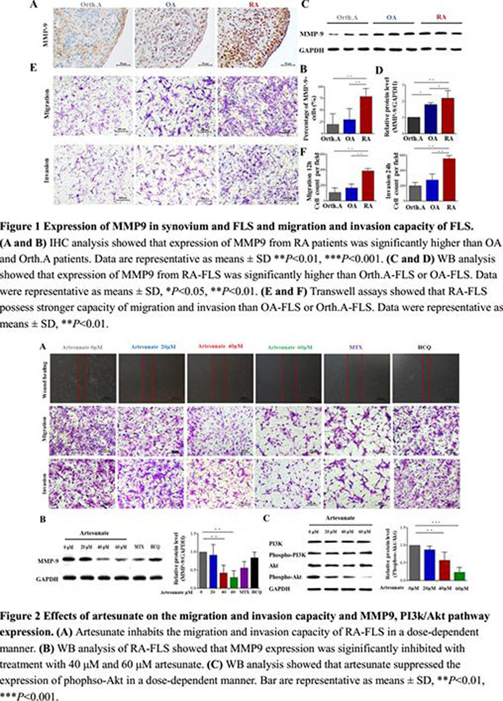
Results: (1) IHC staining showed that synovial MMP9 expressed in lining and sublining area with intense nuclear and endochylema staining in RA synovium and the percentage of MMP9+ cells was significantly higher in RA (n=32) than that in OA (n=6) or Orth.A (n=6, Figure 1A, B).
(2) Migration and wound healing assays for 12 hours and invasion assay for 24 hours showed that RA-FLS possessed stronger capacity in migration and invasion than OA-FLS or Orth.A-FLS (Figure 1E, F). Artesunate inhabits the migration and invasion capacity of RA-FLS in a dose-dependent manner. MTX also has an inhibition effect on the migration and invasion of RA-FLS, but not HCQ (Figure 2A).
(3) MMP9 expression in RA-FLS was significantly higher than that in OA-FLS or Orth.A-FLS (Figure 1C, D). 40μM or 60μM artesunate markedly inhibited the expression of MMP9 in RA-FLS by WB (Figure 2B).
(4) WB analysis showed artesunate suppressed generation of phophso-Akt in a dose-dependent manner which indicated that Akt activity (phophso-Akt/Akt) in 40μM and 60μM artesunate treatment groups were significantly lower than that in untreated group (Figure 2C).
Conclusions: Artesunate could inhibit the migration and invasion capacity of RA-FLS and the expression of MMP9 through suppressing Akt activity.
Acknowledgements: This work was supported by National Natural Science Foundation of China (81671612 and 81471597), Research Project of Traditional Chinese Medicine Bureau of Guangdong Province (20161058) and Guangdong Natural Science Foundation (2014A030313074).
Disclosure of Interest: None declared
DOI: 10.1136/annrheumdis-2017-eular.2039