fetching data ... 
Background: Scleroderma (SSc) is an autoimmune connective tissue disease involving complex interactions between various cell types leading to organ-based tissue fibrosis. Emergence of the monocytes (Mo)/macrophages (Mφ) lineage(s) as key contributors to inflammation, vascular dysfunction and scarring in scleroderma1,2 have led to increased scrutiny of their phenotype and function.
Objectives: To determine the circulating Mo subpopulations and phenotypes of Mφ in SSc.
Methods: PBMC were collected from healthy (HC) and SSc donors, and analysed by flow cytometry using Mo phenotypic antibodies or purified and cultured in vitro. For flow cytometry immunophenotyping, Mo were gated on CD3-CD19-CD56-HLA-DR+populations, and subsets defined by CD14, CD16, CD163 and CD206 expression. For Mφ cultures, Mo were negatively selected from PBMCs, cultured for 7 days, and treated with IFN-γ(5 ng/ml) or IL-4(20 ng/ml) for 24 hours. Cytokine levels in the conditioned media were evaluated by MSD analyses and normalised to total protein levels.
Results: The frequency of circulating CD163+ non-classical Mo (CD14loCD16hi) was 2-fold higher in SSc patients than in HC (unpaired t-test, p=0.026). No difference was found in the frequency of CD206+ monocyte subsets between HC and SSc. In vitro, unstimulated SSc Mφ (M0) secreted higher levels of classically-activated pro-inflammatory (M1) and alternatively-activated pro-regenerative (M2) cytokines. Compared to HC cells, SSc Mφ were more readily polarised towards an M1 phenotype or an M2 phenotype, when cultured in the presence of IFN-γ or IL-4, respectively. Th17 markers and MMPs were significantly increased in SSc Mφ (table 2).
Mo (flow cytometry) |
Mφ supernatant (cytokine assay) |
|||
|---|---|---|---|---|
n |
HC n=9 |
SSc n=10 |
HC n=13 |
SSc n=27 |
Age (years) |
56.7±14.3 |
50.7±5.7 |
60.6±16.7 |
52.1±13.0 |
Female : Male |
7:2 |
8:2 |
6:7 |
26:5 |
SSc subtype |
- |
dcSSc(10 |
- |
dcSSc(27 |
Disease duration |
- |
≤5 years(10 |
- |
≤5 years(,15>5 year(12 |
Abstract FRI0406 – Table 2 Cytokines significantly increased in SSc vs control. Unpaired t-tests, *p<0.05, **P<0.01.
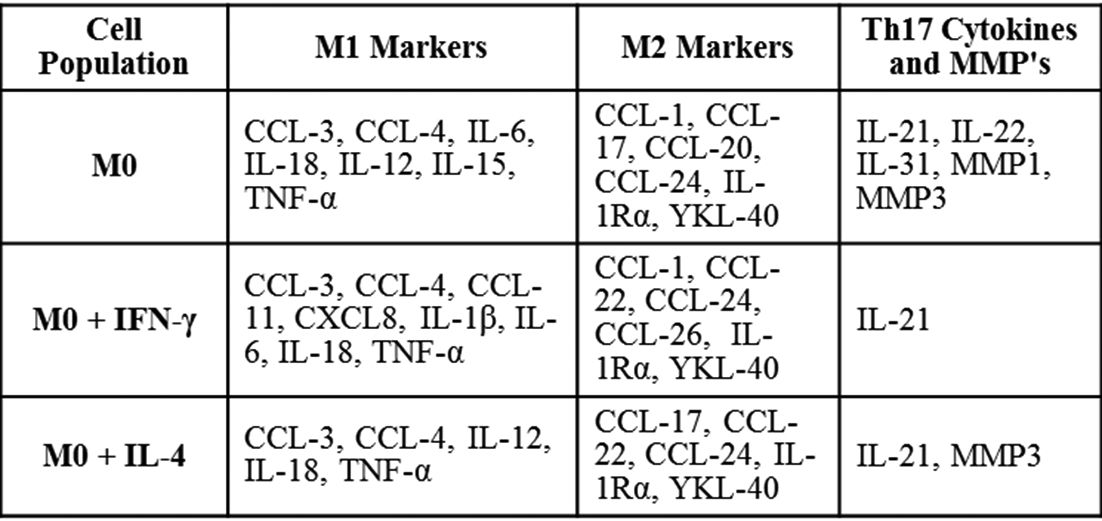
Conclusions: Studies exploring Mo have revealed distinct populations with selective biological functions. Our observation of an increased number of CD163+ non-classical Mo in SSc suggests that this subpopulation may play a key role in inflammatory-driven fibrosis and act as an important source of pro-fibrotic cytokines. This data is consistent with previous reports of elevated serum levels of CD163 and increased CD163 secretion by SSc PBMCs3. SSc Mφ showed a pronounced and enhanced dual M1 and M2 polarisation basally compared to HC, indicating cells were ‘primed’ to undergo phenotypic polarisation. Our studies support the notion that Mφ cytokine secretion generates a pro-fibrotic milieu in scleroderma tissues, playing a prominent role in dysregulated tissue repair in fibrosis.
References:
Disclosure of Interest: None declared
DOI: 10.1136/annrheumdis-2018-eular.1818