fetching data ... 
Background: The study of synovial tissue in patients with Rheumatoid Arthritis (RA) has led to the identification of synovial patterns of immune cell infiltration and specific cellular subsets associated that have been disease activity and clinical outcomes( 1 – 3 ). However, the relationship of circulating and synovial immune cell sub-sets with histopathological features and clinical outcomes remains to be defined.
Objectives: To assess the relationship of peripheral blood and synovial immune cells with RA histopathology and clinical outcomes, by performing flow and digital cytometry in matched peripheral blood and synovial samples from patients with early RA.
Methods: 70 patients with early (<12 months) untreated RA (2010 criteria) recruited in the pathobiology of early Arthritis Cohort (PEAC) at the Barts Health NHS Trust were included( 1 ). Peripheral blood mononuclear cells (n=70) were analysed by flow cytometry. Matched synovial tissues (n=70) obtained by minimally invasive ultrasound-guided synovial biopsy underwent semi-quantitative scoring (0-4) of immune cell infiltration and classification into lympho-myeloid (LM), diffuse-myeloid (DM) and pauci-immune (PI) pathotypes, as previously described( 1 ). 49 synovial and 36 matched peripheral blood samples underwent RNA-sequencing and were analysed by digital cytometry (Xcell) ( 4 ) and Singular Value Decomposition (SVD).
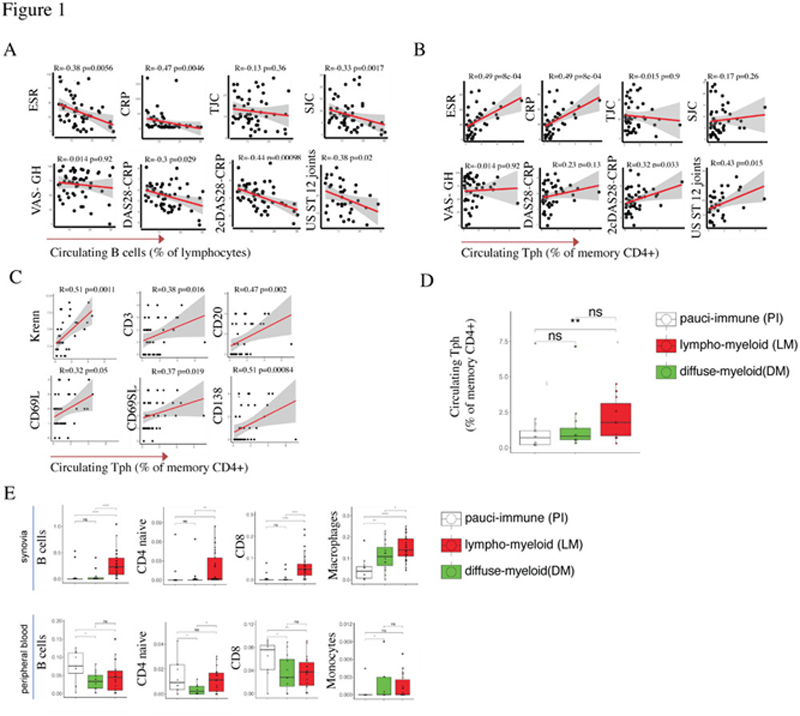
Results: Circulating B cells and their subsets showed significant inverse correlations with inflammatory markers (ESR, CRP), disease activity (swollen joints, four components and two components(
5
) DAS28) and ultrasound scores (
Conclusion: By combining conventional flow cytometry in the peripheral blood and digital cytometry on matched synovial and peripheral blood samples, we highlight diverging associations of circulating immune cell subsets with synovial inflammation and pathotypes. Tph cells, in particular, emerge as predictors of lympho-myeloid synovial inflammation and disease progression.
REFERENCES:
[1]F. Humby et al. , Ann. Rheum. Dis. 78, 761–772 (2019), doi:10.1136/annrheumdis-2018-214539. [2]M. J. Lewis et al. , Cell Rep. 28, 2455-2470.e5 (2019), doi:10.1016/j.celrep.2019.07.091.[3]D. a Rao et al. , Nat. Publ. Gr. 542, 110–114 (2017), doi:10.1038/nature20810.[4]D. Aran et al. , Genome Biol. 18, 220 (2017), doi:10.1186/s13059-017-1349-1.[5]E. M. A. Hensor et al. , Rheumatology . 58, 1400–1409 (2019), doi:10.1093/rheumatology/kez049.
Acknowledgements: The Pathobiology of Early Arthritis Cohort (PEAC) was supported by the MRC (grant 36661). Versus Arthritis provided funding infrastructure support (grant 20022). F. Rivellese is funded by an NIHR Transitional Research Fellowship (TRF-2018-11-ST2-002). We would like to thank the patients and the clinical and laboratory team (core team) at Queen Mary University of London.
Disclosure of Interests: None declared