fetching data ... 
Background: Large-vessel vasculitides comprise Takayasu arteritis (TA)and giant cell arteritis (GCA). Arterial stenosis and dilatation directly affect prognosis but the mechanism(s) underlying remodeling of the vessel wall have not been identified. Microvesicles (MVs) are membrane-enclosed extracellular vesicles released upon cellular activation and stress and as a consequence of environmental inflammation. MVs maintain features and constituents of their parental cells. They have been proposed to serve as potential liquid biopsies in oncology.
Objectives: To verify whether arterial wall derived-MVs are recognizable in the blood of TA patients and express bioactive molecules potentially involved in arterial injury, inflammation and remodeling.
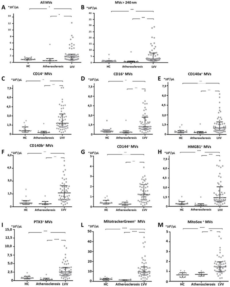
Methods: Platelet was obtained from 112 LVV pts (73 TA, 39 GCA), 42 age and age- and sex-matched healthy controls (HC) and 30 pts with severe carotid atherosclerosis requiring vascular surgery. Plasma flow cytometry was performed with anti-CD14, CD16, anti-CD144 (VE-cadherin, an endothelial marker), anti-CD140a/b (PDGF receptor A/B a vascular stromal marker), anti-HMGB1, anti-PTX3, mitotracker green (that identifies mithochondrial moieties) and mitosox (that revels mitochondrial reactive oxygen species). MVs were identified by physical parameters using Gigamix beads. Medium- to large-sized MVs were defined as MVs with >240nm-eq diameter.
Results: Preliminary results are available for 49 LVV (42 TA, 7 GCA), 8 severe carotidatherosclerosis and 14 age- and sex-matched HC. As compared to HC or CA, LVV plasma contains a higher number of MVs and in particular of medium- to large- sized MVs (p<0.001 for all comparisons) (
Conclusion: MVs, including those expressing arterial stromal biomarkers, are increased in LVV plasma, suggesting a communication between the vessel wall and peripheral blood. MV express signals that may in turn contribute to persisting vascular inflammation in large vessel vascultis Further analysis is required to dissect their potential use as disease biomarkers
REFERENCES:
[1]van Niel G et al, Nat Rev Mol Cell Biol. 2018
[2]Mason JC. Nat Rev Rheumatol. 2010
Disclosure of Interests: None declared