fetching data ... 
Background: AI-enabled algorithms can increase the speed and accuracy of identifying key histological features and enable researchers and clinicians to more readily and thoroughly understand the tissue collected from their patients. Sjogren’s Disease (SJD), in particular, is ripe with opportunity given the reliance on tissue reads for focus scores and overall histological examinations of patients.
Objectives: To develop an AI-enabled algorithm that automatically identifies key histological features of the minor salivary gland of SJD patients, and then test this algorithm on samples from a diverse patient population.
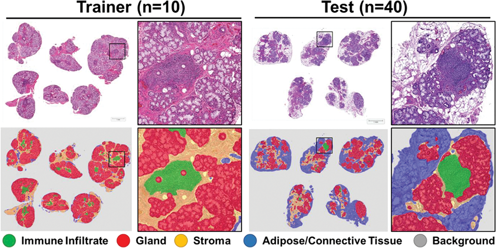
Methods: Minor salivary glands from control and SJD patients were collected via standard-of-care and formalin fixed and paraffin embedded. 5micron sections were collected from blocks containing 3-5 minor salivary glands from a single patient, stained with H&E, then imaged on a whole-slide scanner. Images were loaded into HALO-AI v4.0 (Indica Labs). 5 cases and 5 controls were fully annotated under the guidance of a trained pathologist for background (no tissue), adipose/connective tissue, stroma, glandular tissue, and immune infiltrates. The classes were trained via HALO-AI’s DenseNet v2 for >10,000 iterations with a final entropy of <1 (a measure of agreement between annotation input and AI-prediction). After successful training and implementation on the training set on 10 cases/controls, the algorithm was applied to a set of 40 cases to evaluate performance (n=50 total). Percent area of all classes were then calculated.
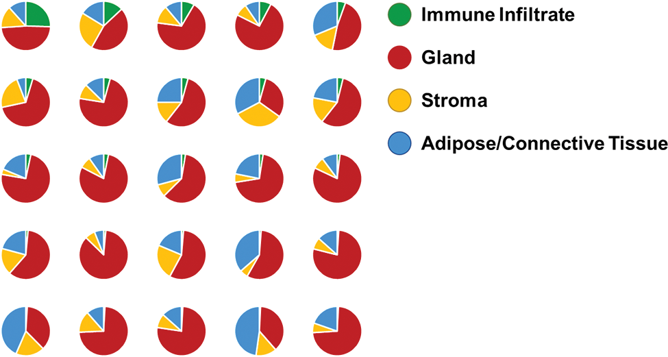
Results: The algorithm accurately identifies all 5 classes across varying degrees of disease severity across SJD patients and controls, as well as differences in staining intensity. Though inaccuracies are observed, the overall results were better than previous machine-learning methodologies (data not shown/published) and were quickly applied to samples (approximately 30sec of analysis time per sample). Resulting data show a high degree in variability in percent immune infiltration and gland.
Conclusion: Though pathologist reads are important for the understanding SjD and clinical workups, it is time consuming and costly to annotate entire images manually in order to measure all major histological features in the minor salivary gland of SJD patients. Though the AI-Tissue classifier produced here would not outperform a trained pathologist in accuracy, it is much faster and much more cost effective to run. Therefore, this algorithm in combination with current focus scores (and other clinical data) could provide novel insights and key findings for both clinicians and researchers concerning SJD progression, severity, or other meaningful metrics.
REFERENCES: NIL.
AI-Tissue Classifier Results of Example Trainer and Test Images.
Percent Histological Features as Identified by the AI-Tissue Classifier and Stratified by Percent Immune Infiltration (High to low, top 50% of immune infiltrate)
Acknowledgements: NIL.
Disclosure of Interests: None declared.