fetching data ... 
Background: Giant cell arteritis (GCA) is a large vessel vasculitis characterized by arterial wall inflammation and remodeling. Highly organized B-cell clusters are present in GCA-affected arteries, particularly in the adventitia of aorta [1]. Fibroblasts, the predominant cell type in the adventitia, may interact with B-cells owing to their location and phenotypic plasticity. We previously showed that in GCA arterial lesions, both B-cells and fibroblasts produce cytokines.
Objectives: This study aims to explore the subtypes of fibroblast in B-cell clusters of GCA-affected aorta tissues, and to investigate the impact of B-cells on fibroblast activation and cytokine production through in vitro analyses.
Methods: Immunohistochemistry, detecting several fibroblast markers (CD90, fibroblast activation protein alpha (FAP), podoplanin (PDPN), CD248, alpha-smooth muscle actin (α-SMA)) and B-cells (CD20), was performed on GCA-affected (n=9) and atherosclerotic (AS, n=11) aorta tissues. B-cells isolated from peripheral blood mononuclear cells (PBMCs) of GCA patients (n=10) and healthy donors (n=10), were incubated with/without CpG oligodeoxynucleotides (ODN) 2006, phorbol 12-myristate 13-acetate (PMA) and calcium ionophore (CaI) for 72 hours. Human aortic adventitial fibroblasts were then cultured with B-cells-conditioned medium or with B-cells for 24 hours in either a co-culture or transwell system. Interleukin(IL)-6 levels in culture medium were measured by ELISA. RNA expression of IL-6, FAP, PDPN, granulocyte macrophage colony-stimulating factor (GM-CSF) and α-SMA in fibroblasts was examined by qPCR.
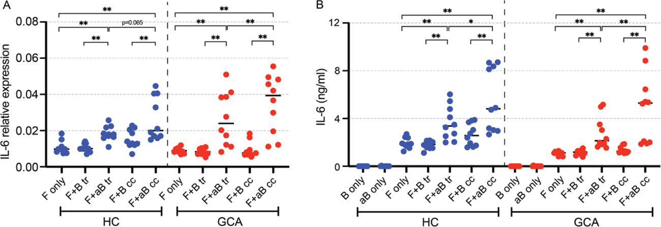
Results: Abundant expression of CD90, PDPN, and CD248 was observed in the B cell clusters in adventitia, with no significant differences in fibroblast phenotypes between GCA-affected aorta and AS aorta. Fibroblasts showed enhanced RNA expression of IL-6 (Figure 1A), FAP, PDPN and GM-CSF, but not α-SMA, when cocultured with activated B-cells or activated B-cells-conditioned medium. Coculture with activated B-cells also promoted IL-6 protein secretion by fibroblasts (Figure 1B). Mechanistically, the effect of activated B-cells on fibroblasts was mainly mediated by soluble factors, and further boosted by cell-cell contact.
Conclusion: Activated B-cells steer fibroblasts towards a proinflammatory phenotype in vitro, possibly contributing to disease chronicity and progression in GCA. If proven, B-cell-targeted therapy may alleviate both B-cell and fibroblasts-related inflammation in GCA.
Activated B-cells promote interleukin-6 (IL-6) expression and secretion in human aortic adventitial fibroblasts. (A) IL-6 mRNA levels (relative to GAPDH) in fibroblasts, cultured with/without B-cells from healthy donors (HC) or GCA patients. (B) IL-6 levels in coculture medium. Abbreviations: GCA=giant cell arteritis, F=fibroblasts, B=B-cells, aB=activated B-cells, tr=transwell, cc=cell cell contact. *p<0.05, **p<0.01.
REFERENCES: [1] Graver JC, et al. Front Immunol. 2019; 10: 83.
Acknowledgements: NIL.
Disclosure of Interests: None declared.