fetching data ... 
Background: Interstitial lung disease (ILD) has been described in approximately 20% of patients with primary Sjogren’s syndrome (pSS), although the demographic, serological and radiological characteristics of these patients remain poorly studied.
Objectives: to evaluate and describe demographic, clinical and serological features of pSS-ILD regularly followed in Italian Rheumatology Units with experience in multidisciplinary team for rheumatologic patients with ILD (mainly pulmonologist, radiologist, rheumatologist).
Methods: One-hundred and thirty-nine pSS ILD patients from 10 Italian rheumatology centres, were enrolled in a cross-sectional study. All patients satisfied current classification criteria for pSS. Diagnosis of ILD was performed by high resolution computed tomography and radiologic patterns were classified according to criteria defined in White Paper of Fleischner Society.
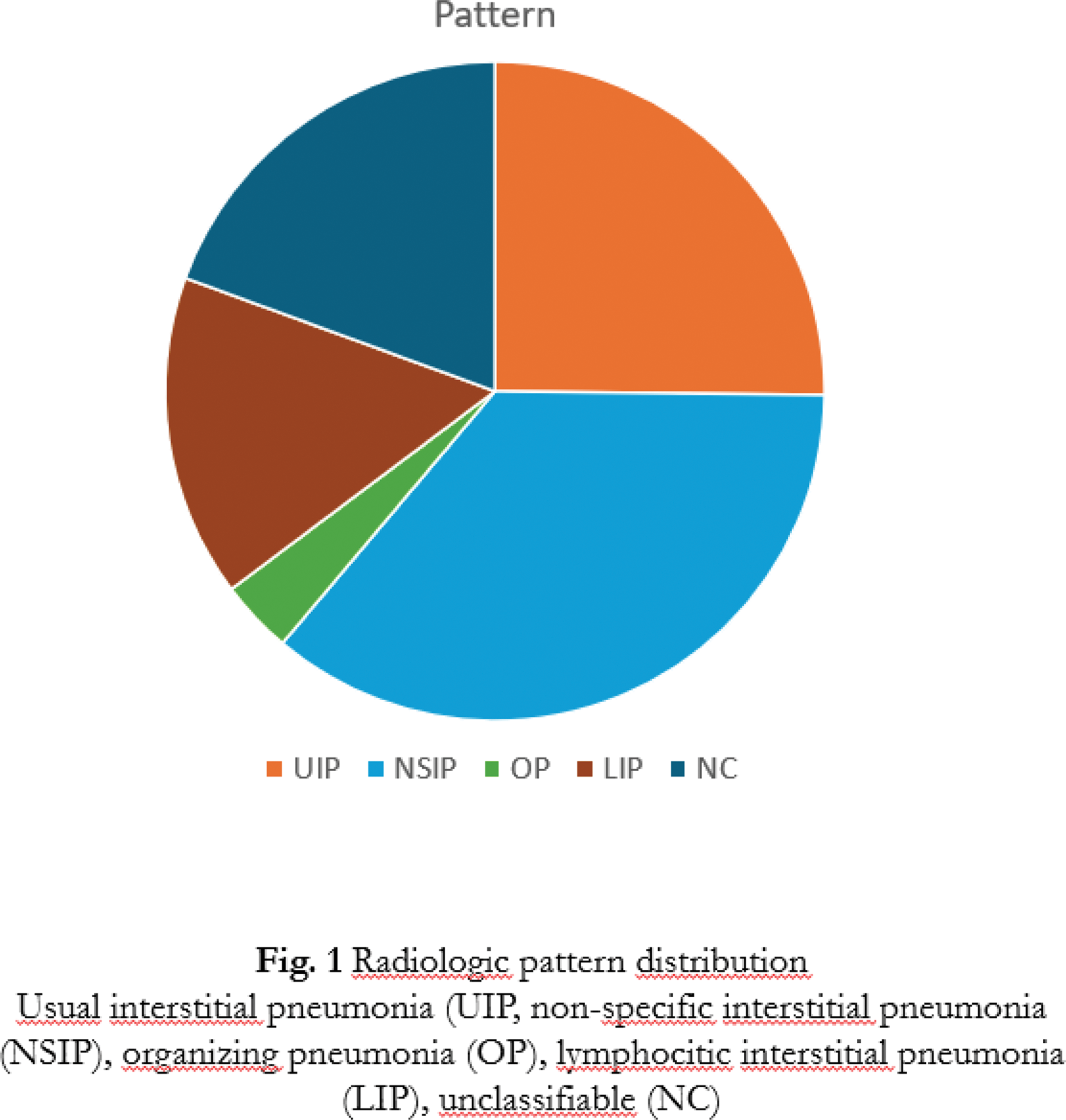
Results: Among patients enrolled, 23 were males and 116 females, mean age at diagnosis of pSS was 60.4±15.3 years, while mean diseases duration of pSS at diagnosis of ILD was 3.7±9.3 years. Smoking habit was detected in 37 patients (26.7%), antinuclear antibodies (ANA) were positive in 115 (83.4%); among them, anti-SSA were detected in 84.3% of cases (97 patients), with positivity for anti-Ro-52 and Ro-60, respectively in 74 (76.3%) and 68 (70.1%) of patients with anti-SSA positivity. Rheumatoid factor was positive in 39.6 % of cases. Mean forced vital capacity (FVC) showed normal value (94,5±22,5% of predicted) while lung diffusion test (DLCO) was moderately reduced (60,8±18,3% of predicted). Diagnosis of ILD preceded or was concurrent with diagnosis oof pSS in 64 patients (46% of cases). From a radiologic point of view, a definite or probable usual interstitial pneumonia (UIP) pattern was observed in 35 patients (25.2%), a nonspecific interstitial pneumonia (NSIP) pattern was observed in 50 (35.9%), an organising pneumonia in 5 (3.6%), while a lymphocytic interstitial pneumonia was detected in 22 (15.8%). In 27 patients the pattern was not classifiable (Figure 1). A UIP pattern was detected in 34.8% of patients in which ILD was diagnosed before or concurrently with pSS diagnosis, other radiologic diagnosis for this group were NSIP in 36.3%, and LIP in 7.6%. In patients in which ILD was diagnosed in the course of pSS radiologic classification was UIP pattern in 16.4% of patients, NSIP pattern in 37.4%, and LIP in 25.4%.
Conclusion: In a large case series of patients with pSS, our data show that the timing of onset of ILD is associated with different radiological features. Indeed, a prevalence of fibrosing patterns, and in particular the UIP pattern, is observed in patients with early-onset ILD.
REFERENCES: [1] Luppi F, Sebastiani M, Sverzellati N, Cavazza A, Salvarani C, Manfredi A. Lung complications of Sjogren syndrome. Eur Respir Rev. 2020;29:200021.
Acknowledgements: NIL.
Disclosure of Interests: None declared.
© The Authors 2025. This abstract is an open access article published in Annals of Rheumatic Diseases under the CC BY-NC-ND license (