fetching data ... 
Background: Cutaneous lupus erythematosus (CLE) is a frequent and clinically relevant manifestation of lupus erythematosus, occurring either as an isolated condition or in association with systemic disease. Differential diagnosis is often challenging due to significant clinical overlap with other inflammatory dermatoses, particularly rosacea in facial involvement. Although histopathology remains the diagnostic gold standard, skin biopsy is invasive, may cause scarring, and is poorly suited for repeated assessments [1-3]. Line-Field Confocal Optical Coherence Tomography (LC-OCT) is a novel non-invasive imaging technique that enables real-time, in vivo visualization of the skin with near-histological resolution to a depth of approximately 500 μm. While LC-OCT has shown diagnostic potential in neoplastic and inflammatory skin diseases, its role in CLE remains poorly studied, with the limited available evidence mainly focusing on the discoid variant [4-5]. Its performance across different CLE subtypes and in clinically relevant differential diagnosis are limited.
Objectives: The aim of this pilot study is to characterize LC-OCT features of acute (ACLE), subacute (SCLE), and chronic (CCLE) cutaneous lupus erythematosus and to explore whether LC-OCT identifies distinct in vivo patterns between ACLE and rosacea.
Methods: This cross-sectional, single-center observational study was conducted between November 2023 and September 2025. Consecutive adult patients with CLE lesions (ACLE, SCLE, CCLE) and a comparator group with active rosacea were enrolled. All participants underwent standardized clinical and LC-OCT imaging (DeepLive™, DAMAE Medical). Multiple vertical B-scans, one video scan, and a 3D stack were acquired from representative lesions; adjacent unaffected skin served as control. Two blinded, LC-OCT–trained dermatologists independently evaluated predefined morphological criteria corresponding to known histopathologic features of CLE. Interobserver agreement was assessed using Cohen’s κ. Exploratory comparisons between ACLE malar rash and rosacea were performed using Fisher’s exact test with Benjamini–Hochberg correction.
Results: 45 participants were included: 30 with CLE (16 ACLE, 5 SCLE, 9 CCLE), including 14 patients with systemic lupus erythematosus (SLE), and 15 with rosacea.
LC-OCT revealed layer-specific and stage-dependent morphological signatures in CLE lesions:
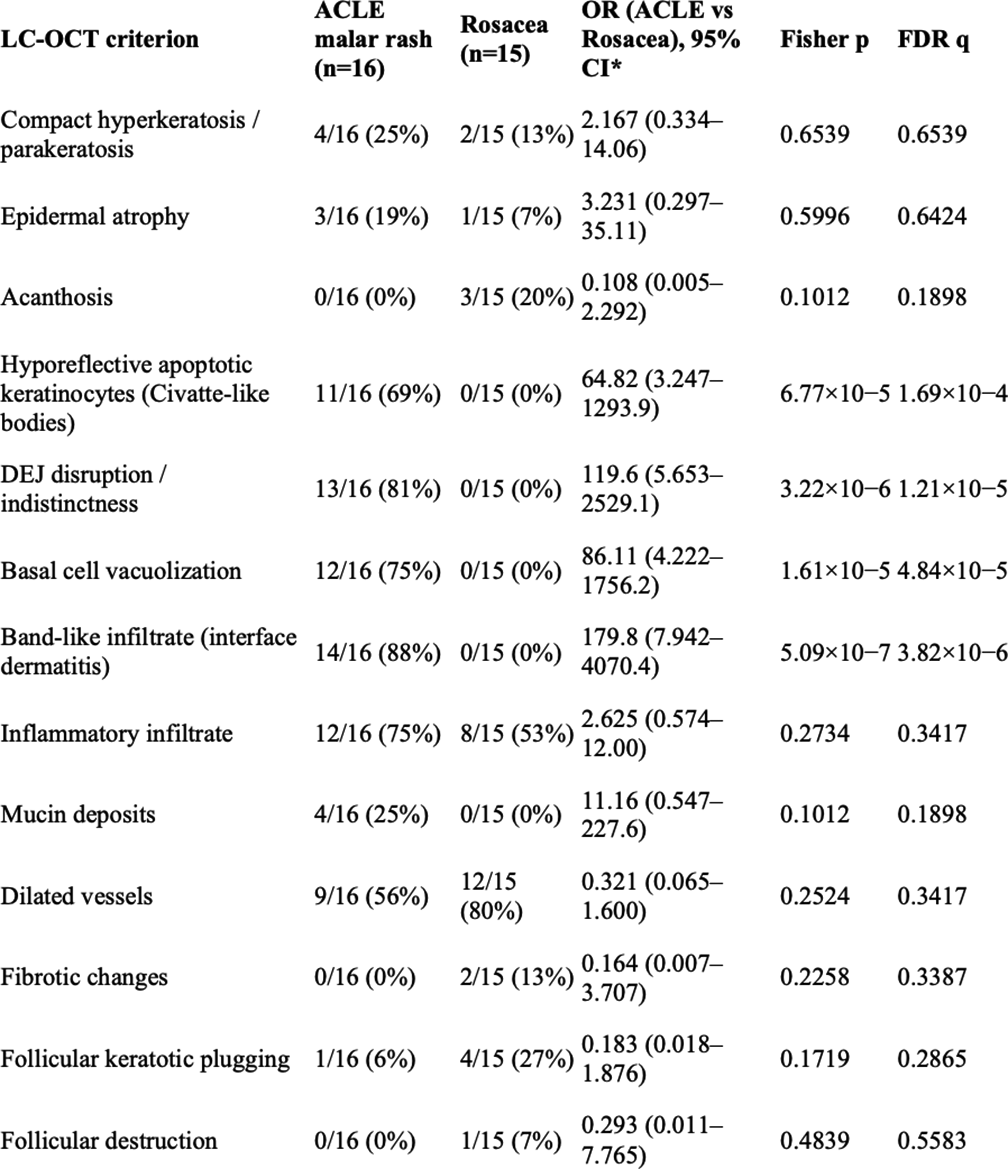
- Epidermal findings: Compact hyperkeratosis/parakeratosis was most frequent in CCLE (89%), intermediate in SCLE (60%), and limited in ACLE (25%). Epidermal atrophy followed a similar pattern (CCLE 78%, SCLE 40%, ACLE 19%). Hyporeflective apoptotic keratinocytes (“Civatte-like bodies”) were enriched in ACLE/SCLE (69/60%) vs CCLE (33%). - DEJ/interface changes: DEJ disruption (ACLE 81%, SCLE 80%, CCLE 56%), basal cell vacuolization (75%, 60%, 44%) and band-like interface infiltrate (88%, 80%, 56%) were consistently observed in active lesions and absent in non-lesional skin. - Dermal alterations: Inflammatory infiltrate was present in 60–78% of CLE lesions; mucin deposits in 25–40%; dermal fibrosis—recognized as parallelized hyperreflective collagen bundles—was the hallmark of CCLE (89%). - Adnexal involvement: Follicular keratotic plugging (89%) and follicular destruction (67%) characterized CCLE, while peri-infundibular infiltrates were observed in 78%. Overall, fibrotic remodeling and adnexal destruction were absent in ACLE but common in CCLE, reflecting chronicity. Interobserver agreement was substantial to excellent (κ=0.80–1.00 for most structural features; lowest for mucin κ=0.53). In the ACLE versus rosacea comparison, LC-OCT revealed clearly divergent patterns. Interface dermatitis features—band-like DEJ infiltrate (88% vs 0%), DEJ disruption (81% vs 0%), basal vacuolization (75% vs 0%), and Civatte-like bodies (69% vs 0%)—were strongly associated with ACLE (all q < 0.05). Conversely, rosacea exhibited a vascular-folliculocentric profile with peri-infundibular infiltrates (100% vs 19%) and Demodex mites (87% vs 0%) (both q < 0.05) (Table1; Figure1).
Conclusions: LC-OCT enables real-time, non-invasive visualization of key histopathologic correlates of CLE, including interface dermatitis, dermal fibrosis, and adnexal remodeling, and highlights distinct morphological differences between ACLE and rosacea through epidermal and DEJ patterns. Moreover, LC-OCT captured in vivo morphological features associated with disease activity, providing additional, layer-specific information beyond clinical inspection. These preliminary findings suggest a potential role for LC-OCT in supporting differential diagnosis, disease monitoring, and in reducing unnecessary biopsies, particularly in cosmetically sensitive areas. Future studies in larger, well-characterized cohorts are needed to integrate site-matched histopathologic comparison, the development of standardized interpretative criteria, finer immunological characterization of SLE patients for correlation with systemic disease activity to further define the role of LC-OCT as a ‘virtual biopsy’ adjunct in CLE, bridging clinical observation and histology.
Representative (LC-OCT) images of ACLE (A), rosacea (B), SCLE (C), and CCLE (D).
In particular, interface dermatitis (ID), hyperkeratosis (HK), a disrupted dermoepidermal junction (DD), dilated vessels (DV), basal keratinocyte apoptosis and vacuolization (white circles), dermal fibrotic changes (DF) and periappendageal inflammatory infiltrate (white arrow) are visible. These features are absent in rosacea, in which a cluster of Demodex folliculorum mites (DF) is instead observed in a follicle.
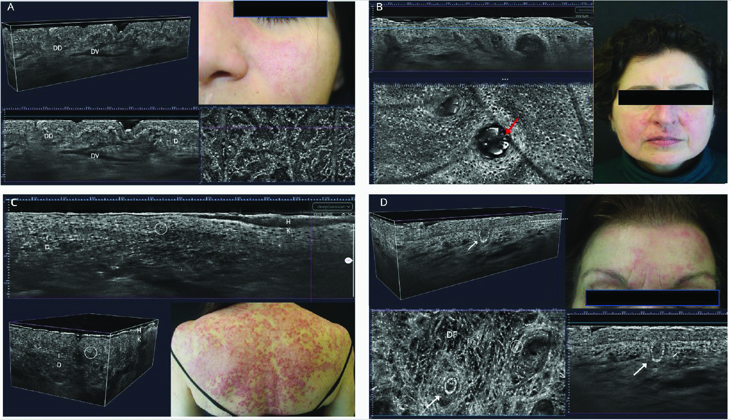
Table 1. Frequency of LC-OCT features in the malar rash, typical of ACLE and rosacea
REFERENCES: [1] Worm M et al. J Dtsch Dermatol Ges. 2021;19(8):1236–1247.
[2] Hobayan CGP et al. Lupus Sci Med. 2024;11(2):e001280.
[3] Brown TT et al. J Am Acad Dermatol. 2014;71(1):100–107.
[4] Benvenuto P et al. Dermatol Pract Concept. 2025;15(1):5089.
[5] Maione V et al. Exp Dermatol. 2025;34(12):e70184.
Acknowledgments: NIL.
Disclosure of Interests: None declared.