fetching data ... 
Background: Imaging is a key tool for timely diagnosis and for monitoring inflammation and structural damage in patients with spondyloarthritis (SpA). The original EULAR recommendations for the use of imaging in the diagnosis and management of SpA in clinical practice reflect the evidence and expert opinion as it was in 2012 [1]. Since then, a large number of pivotal studies have been published in the field, which have changed the way imaging is used in SpA, highlighting the need for an update
Objectives: To update the original evidence-based EULAR recommendations on the use of imaging in the diagnosis and clinical management of both axial (axSpA) and peripheral SpA (pSpA) for clinical practice.
Methods: The task force comprised an expert group of 26 rheumatologists, radiologists, health care professionals, methodologists, patient research partners and members of EMEUNET from 15 EULAR countries. Twelve key research questions on the role of imaging in SpA, originally formulated using the PICO method for the original recommendations, were adopted for the update. Imaging modalities included radiography, ultrasound (US), magnetic resonance imaging (MRI), computed tomography (CT), positron emission- and single photon emission computed tomography, dual-emission x-ray absorptiometry (DXA) and scintigraphy. Experts used research evidence obtained from two systematic literature reviews using MEDLINE, EMBASE and CENTRAL, based on the search strategy developed for the original work, to revise, update and/or create new recommendations. Risk of bias of the included studies was assessed using standardized instruments depending on the study type. Level of evidence (LoE) and grade of recommendation (GoR) were evaluated, level of agreement (LoA), as well as impact, feasibility and priority of the recommendations were assessed by the task force members using the 1-10 Likert scale and rank order, respectively.
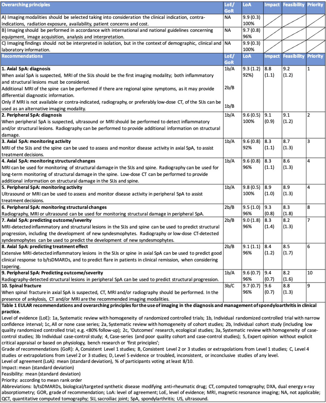
Results: A total of 11,656 references were identified in the search process, and 181 studies were included. The task force formulated three new overarching principles, updated nine of the original recommendations, added one new recommendation, and did not formulate recommendations for two of the research questions. The final recommendations encompass the role of imaging in making a diagnosis of axSpA or pSpA, monitoring inflammation and damage, predicting outcome and response to treatment, and detecting spinal fractures (Table 1). The LoE/GoR ranged from 1-3b and A-C, respectively. The LoA were generally very high (range: 9.0-9.9). Impact and feasibility ranged between 8.3-9.6 and 8.2-9.2 respectively.
The main changes in the update as compared to the original recommendations include replacing radiography with MRI of the sacroiliac joints (SIJs), and additionally of the spine in case of symptoms in that region, as the first imaging modality for diagnosing axSpA. Radiography or preferably low-dose CT, which was not mentioned in the previous recommendations for diagnostic purposes, of the SIJs are suggested as alternative modalities, if MRI is not available or contra-indicated. For diagnosis of pSpA, US and MRI remain the imaging methods of choice, and in addition to inflammatory lesions, structural lesions should now also be considered. For monitoring axSpA, MRI can be used to follow up inflammation and structural damage in the SIJs and spine, while radiography is suitable for long-term monitoring of structural damage in the spine. For monitoring structural damage in pSpA, US and MRI are now recommended along with radiography. In the SIJs or spine in axSpA, MRI-detected inflammatory and structural lesions can be used to predict structural progression, while extensive MRI-detected inflammatory lesions can be used to predict good clinical response to biological/targeted synthetic disease modifying antirheumatic drugs (b/tsDMARDs), and to predict flare in patients in clinical remission. In a new recommendation, structural lesions on radiographs of the peripheral joints are suggested for predicting structural progression in pSpA. Finally, CT, MRI and/or radiography are recommended in case of suspected spinal fracture.
Conclusions: The task force successfully formulated three new overarching principles, updated nine recommendations, and added one new recommendation with regard to imaging using research-based evidence and expert opinion in clinical practice of SpA.
REFERENCES: [1] Mandl P, Navarro-Compán V, Terslev L, et al. European League Against Rheumatism (EULAR). EULAR recommendations for the use of imaging in the diagnosis and management of spondyloarthritis in clinical practice. Ann Rheum Dis. 2015 Jul;74(7):1327-39.
Acknowledgments: NIL.
Disclosure of Interests: Peter Mandl Abbvie, Eli-Lilly, Novartis, Sobi, UCB, AbbVie, Alfasigma, Eli Lilly, Novartis, Sobi, UCB, Clara Watschinger: None declared, Susanne Juhl Pedersen MSD, Pfizer, AbbVie, UCB, Novartis, AbbVie, UCB, Novartis, AbbVie, MSD, Novartis, and the Danish Research Foundation, Delphine S Courvoisier: None declared, Lennart Jans: None declared, Lene Terslev: None declared, Philip G. Conaghan AbbVie, Alfasigma, Eli Lilly, Eupraxia, Formation Bio, Genascence, GSK, Grunenthal, Janssen, Kolon TissueGene, Levicept, Moebius, Novartis, Orion, Pacira & Takeda, Esperanza Naredo AbbVie, Janssen, and Novartis, Eli Lilly and AbbVie, Iwona Sudol-Szopinska: None declared, Anna E F Hadsbjerg: None declared, Manouk de Hooge UCB, Ulrich Weber AbbVie, Novartis, Eli Lilly, Sylvia Taxer: None declared, Annamaria Iagnocco: None declared, Maria-Antonietta D’Agostino: None declared, Iris Eshed: None declared, Torsten Diekhoff: None declared, Russka Shumnalieva AstraZeneca, Novartis, Abbvie, Janssen,, Yeliz Prior: None declared, Kristyna Bubova: None declared, Olivier FAKIH: None declared, Pedro Machado Abbvie, BMS, Celgene, Eli Lilly, Galapagos/Alfasigma, Greywolf, Janssen, MSD, Novartis, Orphazyme, Pfizer, Roche and UCB, Miranda van Lunteren: None declared, Xenofon Baraliakos: None declared, Victoria Navarro-Compán Abbvie, Alfasigma, BMS, Fresenius Kabi, Galapagos, Janssen, Lilly, Moonlake, MSD, Novartis, Pfizer, Roche, UCB, Abbvie, Alfasigma, BMS, Fresenius Kabi, Galapagos, Janssen, Lilly, Moonlake, MSD, Novartis, Pfizer, Roche, UCB, Abbvie, Alfasigma, BMS, Fresenius Kabi, Galapagos, Janssen, Lilly, Moonlake, MSD, Novartis, Pfizer, Roche, UCB, Mikkel Østergaard Abbvie, BMS, Celgene, Eli-Lilly, Galapagos, Gilead, Janssen, MEDAC, Merck, Novartis, Pfizer and UCB, Abbvie, AlfaSigma, BMS, Celgene, Eli-Lilly, Galapagos, Gilead, Janssen, Merck, Novartis, Pfizer, and UCB, AbbVie, Amgen, BMS, Merck, Celgene, Eli Lilly, Novartis, and UCB,.